
Paid online consultations are available by appointment. For booking, please contact us via Whatsapp.

Aneurysms

Table of Contents
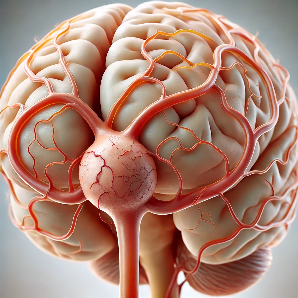
What Are Brain Aneurysms?
Brain aneurysms, also known as cerebral aneurysms, are abnormal bulges in the walls of brain arteries, most commonly found at branching points (bifurcations). Due to their berry-like appearance with a neck and dome, they are also referred to as berry aneurysms. These vascular sacs are dangerous because they can tear (rupture) and cause severe bleeding in the brain, leading to conditions such as subarachnoid hemorrhage, coma, or even death.
Are Brain Aneurysms Common?
It is estimated that 3-5% of the general population may have a brain aneurysm, though the risk of rupture is low, occurring in about 5-10 per 100,000 individuals annually. Many aneurysms remain undiagnosed until they rupture or grow large enough to cause symptoms.
Causes and Risk Factors
While the exact cause of brain aneurysms is unclear, several factors contribute to their development:
- Smoking and drug use (e.g., cocaine) increase the risk of aneurysm formation.
- Hypertension and atherosclerosis can weaken artery walls and promote aneurysm growth.
- Genetics: Family history may slightly increase the risk, raising the question, “Are brain aneurysms hereditary?” Although most aneurysms are not inherited, some genetic conditions may predispose individuals to aneurysm formation.
Types and Symptoms of Brain Aneurysms
Brain aneurysms vary in shape and size:
- Saccular (berry) aneurysms: The most common type.
- Fusiform aneurysms: Bulging along a segment of the artery.
- Dissecting aneurysms: Caused by a tear in the artery wall.
Symptoms often depend on the size and location of the aneurysm:
- Small aneurysms: Often asymptomatic.
- Large aneurysms (over 25 mm): May press on nerves or brain tissue, causing:
- Drooping eyelids (ptosis)
- Headaches
- Blurred vision or difficulty moving the eyes
- Muscle weakness or numbness on one side of the body
- Unruptured Aneurysm
Often asymptomatic unless it presses on surrounding tissues.
Possible symptoms:
Headaches (localized or generalized).
Pain above or behind the eye.
Vision changes, such as blurred or double vision.
Difficulty speaking or understanding speech.
Weakness or numbness in one side of the face or body. - Ruptured Aneurysm
Sudden, severe headache (“thunderclap headache”) — often described as the worst headache of one’s life.
Nausea and vomiting.
Stiff neck.
Sensitivity to light (photophobia).
Loss of consciousness or confusion.
Seizures.
Can Brain Aneurysms Be Detected?
Yes, brain aneurysms can be detected through advanced imaging techniques:
- Computed Tomography Angiography (CTA) and Magnetic Resonance Angiography (MRA) are non-invasive methods for diagnosis.
- Digital Subtraction Angiography (DSA) provides detailed images but requires catheterization.
- In emergencies, a CT scan is often the fastest way to detect bleeding in the brain.
Can Brain Aneurysms Be Prevented?
While it is impossible to entirely prevent brain aneurysms, reducing risk factors can help:
- Avoid smoking and illicit drugs.
- Manage high blood pressure and cholesterol.
- Maintain a healthy lifestyle with regular exercise and a balanced diet.
Are Brain Aneurysms Fatal?
Ruptured brain aneurysms are a medical emergency and can be fatal. However, early detection and treatment significantly improve outcomes.
Treatment Options for Brain Aneurysms
1. Microsurgical Clipping:
- A metal clip is placed at the base of the aneurysm to cut off blood flow.
- Performed by highly trained neurosurgeons, this procedure requires precision.
2. Endovascular Therapy:
- Minimally invasive techniques, such as coiling, use catheters to secure the aneurysm.
3. Bypass Surgery:
- Used in complex cases where direct intervention is not feasible.
Post-surgery, patients are often monitored in an intensive care unit (ICU) to manage complications like vasospasm (narrowing of blood vessels) and hydrocephalus (fluid buildup in the brain).
Frequently Asked Questions
Can brain aneurysms cause memory loss?
Yes, especially if the aneurysm affects areas of the brain responsible for memory or ruptures, leading to brain damage.
Does a brain aneurysm cause headaches?
Persistent or severe headaches may indicate an aneurysm, particularly if it is large or ruptured.
Can brain aneurysms cause seizures or strokes?
Yes, ruptured aneurysms can trigger seizures or strokes due to bleeding and blood vessel damage.
Can children have brain aneurysms?
Although rare, brain aneurysms can occur in children, typically linked to genetic conditions or trauma.
Is brain aneurysm surgery dangerous?
All surgeries carry risks, but advancements in neurosurgery have made treatments for brain aneurysms safer and more effective.
Conclusion
Brain aneurysms are serious conditions that require prompt medical attention. While the risk of rupture is low, early detection through regular health checkups and imaging can save lives. If you experience symptoms like sudden, severe headaches or neurological issues, seek immediate medical care.
Contact Us