
Paid online consultations are available by appointment. For booking, please contact us via Whatsapp.

In short
What Is a Cavernoma?
A cavernoma, or cerebral cavernous malformation (CCM), is a cluster of thin, leaky blood vessels. On MRI, it looks like a tiny mulberry or a piece of popcorn. It is not a tumor or cancer, but it can leak blood into nearby brain.
Blood moves through a cavernoma slowly and under low pressure. As a result, its bleeds usually stay small, unlike aneurysm or AVM bleeds.
Cavernomas affect roughly 1 in 200 to 1 in 600 people, and many never cause symptoms. Most occur alone; however, about 1 in 5 patients has an inherited form.
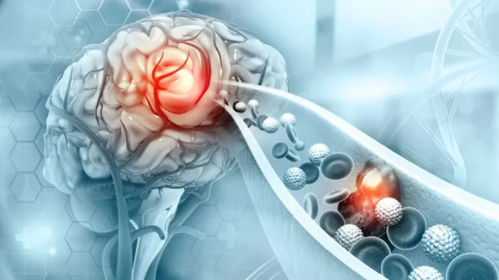
Key facts about cavernomas
A few numbers help put a cavernoma in context. Each one also shapes the treatment decision.
- SizeMost measure about 1 cm; giant ones exceed 3 cm
- LocationMost sit in the cerebral hemispheres; fewer lie in the brainstem
- First symptomSeizures in about half; a bleed or a deficit in the rest
- Multiple lesionsAbout 1 in 5 patients has more than one
- InheritanceAbout 20% have a familial form linked to three genes
- ImagingInvisible on a catheter angiogram; MRI finds it
Which Symptom Brought You Here?
First, pick the change you or your family noticed. Then, each link explains how a cavernoma can cause it.
- 01A seizureThe most common first symptom of a cavernoma
- 02A sudden headache with new weakness or numbnessSigns of a small bleed
- 03Double vision, facial numbness or trouble swallowingTypical of a cavernoma in the brainstem
- 04Several cavernomas or a family historyA possible inherited form
- 05A cavernoma found by chanceNo symptoms, but questions about the future
Cavernoma Symptoms
Up to half of cavernomas never cause symptoms. When they do, the symptom depends on where the cavernoma sits and whether it has bled.
Seizures
Seizures are the first symptom in about half of patients, especially with cavernomas near the brain surface. Small leaks leave iron-rich blood products (hemosiderin), which then irritate nearby brain cells.
Most patients control seizures with medicines. If seizures continue, however, epilepsy surgery principles apply: the surgeon removes the cavernoma and its hemosiderin ring. In suitable patients, this controls seizures in about 70-90% of cases, especially when they started recently.
A small bleed
A bleed causes a sudden headache and new symptoms that match its location. These can include, for instance, weakness, numbness, vision or speech changes.
Many patients recover well after a first bleed. In a large prospective cohort, for example, disability fell from 27% to about 6% within a year.
However, each new bleed makes lasting disability more likely. The risk of yet another bleed also rises for a while.
Brainstem cavernomas
The brainstem packs many nerve pathways into a small space. Therefore, even a small bleed there can cause:
- Double vision
- Facial numbness or weakness
- Trouble swallowing or speaking
- Weakness or numbness of the arms or legs
- Unsteady walking
A brainstem location raises the bleeding risk about fourfold. So these cavernomas need close follow-up by an experienced team.
Several cavernomas or a family history
About 20% of patients have the familial form. It follows autosomal dominant inheritance. So each child of an affected parent has a 50% chance of inheriting it.
Three genes cause it: KRIT1 (CCM1), CCM2 and PDCD10 (CCM3). Generally, PDCD10 variants cause the most severe course, and new cavernomas can appear throughout life.
Genetic testing therefore makes sense with several cavernomas and no venous anomaly, or with affected relatives.
A cavernoma found by chance
More cavernomas now turn up on MRI scans done for other reasons. In fact, their bleeding risk stays low: about 0.08% per patient-year for a first bleed in one study.
Most need only observation. For example, the AANS suggests a yearly MRI for two years, then one every five years. New symptoms, however, call for an earlier scan.
Is a cavernoma a tumor?
No. A cavernoma is a vascular malformation, not a growth of dividing cells. However, repeated small bleeds can make it look larger over time, much like a slowly growing mass.
What is a developmental venous anomaly?
A developmental venous anomaly (DVA) is an unusual but normal vein pattern. It often sits next to a sporadic cavernoma. Surgeons therefore leave it in place, because it drains healthy brain.
Questions at this stage
Can a cavernoma cause headaches or dizziness?
Yes, especially after a bleed or when it sits in the brainstem or cerebellum. However, many headaches have other causes, so doctors look for a match between symptom and location.
Can cavernomas grow or disappear?
They can grow slightly with repeated small bleeds, and new ones can appear in the familial form. However, they do not disappear on their own.
Can I take aspirin or ibuprofen with a cavernoma?
Most studies suggest that antiplatelet medicines such as aspirin carry little risk. Small series also found anti-inflammatory painkillers safe. Still, discuss any blood thinner with your doctor, especially after a recent bleed.
When to See a Doctor
Many cavernomas never cause trouble. However, some signs need a prompt check, and a few need emergency care.
Book a doctor's visit if you notice:
- Headaches that change or get worse
- Seizures that break through despite medicine
- Several cavernomas on an MRI report
- A relative with cavernomas
- A planned pregnancy with a known cavernoma
Seek emergency care for:
- A first seizure, or a seizure lasting over 5 minutes
- A sudden headache with new weakness or numbness
- New double vision, facial numbness or trouble swallowing
- Sudden unsteadiness or slurred speech
After any sudden new symptom, an MRI within days shows whether the cavernoma bled. A CT helps in the first days, but MRI with blood-sensitive sequences remains the key test.
Cavernoma vs AVM: What Is the Difference?
Both are vascular malformations, yet they behave very differently. So the distinction changes the risk, the tests and the treatment.
| Feature | Cavernoma | Brain AVM |
|---|---|---|
| Structure | Small blood-filled caverns with no brain tissue between them | A tangle of arteries and veins without capillaries |
| Blood flow | Very slow, low pressure | Fast, high pressure |
| On a catheter angiogram | Invisible | Clearly visible |
| Best test | MRI with susceptibility-weighted or gradient-echo images | Catheter angiogram plus MRI |
| Typical first sign | A seizure | A bleed or a seizure |
| Bleeding | Usually small; higher risk after a first bleed and in the brainstem | Can be large; about 3% a year on average |
| Inherited form | About 20% (KRIT1, CCM2, PDCD10) | Rare, mainly with HHT |
| Main treatment | Observation or surgery; radiosurgery only in select cases | Surgery, embolization, radiosurgery or observation |
For details, see brain AVMs. Also compare brain aneurysms and dural arteriovenous fistulas, which people often confuse with cavernomas.
Is a cavernoma more dangerous than an AVM?
Usually less, because low pressure keeps cavernoma bleeds small. However, a cavernoma in the brainstem can cause serious deficits even with a small bleed.
Cavernoma Bleeding Risk: What the Numbers Show
Two factors predict a future bleed best: the way the cavernoma first appeared and its location. In contrast, age, sex and the number of cavernomas add little.
| Group | 5-year risk of a symptomatic bleed |
|---|---|
| Outside the brainstem, found without a bleed or deficit | 3.8% |
| In the brainstem, found without a bleed or deficit | 8.0% |
| Outside the brainstem, first showed with a bleed or deficit | 18.4% |
| In the brainstem, first showed with a bleed or deficit | 30.8% |
These figures come from a meta-analysis of 1,620 untreated patients. Moreover, the risk of another bleed tends to fall as time passes after a bleed.
Over 10 years, a 2026 cohort found a disability risk under 8% outside the brainstem. For brainstem cavernomas, in contrast, it reached about 35%.
Cavernoma Treatment: Observation, Surgery or Radiosurgery?
Most cavernomas need only observation. Therefore, surgery makes sense when living with the cavernoma carries more risk than removing it.
| Situation | Usual recommendation |
|---|---|
| Found by chance, no symptoms | Observation with MRI; surgery generally not advised, especially in deep or brainstem locations |
| Seizures controlled with medicine | Observation, or elective surgery for an easily reached cavernoma |
| Seizures despite medicine | Early surgery that removes the cavernoma and its hemosiderin ring |
| Symptoms from an easily reached cavernoma | Surgery, with a risk similar to about 2 years of living with it |
| Deep cavernoma after a bleed (basal ganglia, thalamus, insula) | Surgery in selected cases, with a risk similar to 5-10 years of natural risk |
| Brainstem cavernoma after a second bleed | Surgery is reasonable at an experienced center |
| Bleeding cavernoma where surgery is too risky | Radiosurgery may be considered |
| Familial, multiple cavernomas | Treat only the one causing symptoms; avoid radiosurgery |
How does cavernoma surgery work?
The surgeon reaches the cavernoma through a craniotomy, guided by neuronavigation. For deep or brainstem cavernomas, moreover, tractography and nerve monitoring help choose a safe entry point.
The whole cavernoma must come out, because a leftover piece can bleed again. Meanwhile, the surgeon spares the nearby venous anomaly. For seizures, the surgeon also removes the stained rim of brain around the cavernoma.
Complete removal cures that cavernoma, and it rarely returns. However, people with the familial form can still develop new ones elsewhere.
What about radiosurgery?
Radiosurgery does not remove a cavernoma. Some studies saw fewer bleeds two years after treatment. However, this may simply mirror the natural fall in risk over time.
It also carries real risks: a meta-analysis of brainstem cases reported 5.6% mortality and 11.8% new deficits. Therefore, guidelines reserve it for bleeding cavernomas in areas too risky for surgery. They also advise against it in the familial form, because radiation may trigger new cavernomas.
What is the success rate of cavernoma surgery?
Complete removal cures the treated cavernoma and removes its bleeding risk. For epilepsy, about 70-90% of well-selected patients gain seizure control. Risks, however, depend mainly on location.
Your treatment path
From Diagnosis to Recovery
Care follows four steps. The MRI, especially its blood-sensitive sequences, guides each of them.
STEP 01
Diagnosis: the right MRI
On MRI, a cavernoma shows a speckled "popcorn" core with a dark rim of old blood. Susceptibility-weighted (SWI) or gradient-echo sequences also reveal tiny cavernomas that standard images miss.
- CT helps in the first days after a bleed, but it can miss small cavernomas.
- A catheter angiogram usually adds nothing, because cavernomas stay invisible on it.
- Genetic testing clarifies the familial form when several cavernomas appear.
Why did my CT look normal if I have a cavernoma?
Small cavernomas often hide on CT. MRI, especially with blood-sensitive sequences, detects them with near-perfect sensitivity.
STEP 02
Planning: when and how
Next, the team weighs location, symptoms and bleeding history against the risk of surgery. For deep cavernomas, tractography and functional MRI map the nearby pathways for movement and speech.
For brainstem cavernomas, timing also matters. Surgeons often operate in the weeks after a bleed. By then, the lesion may reach the surface, or the clot may open a safer path.
STEP 03
Surgery: complete removal
The operation takes place under general anesthesia, with nerve monitoring for deep or brainstem lesions. Then, the surgeon removes the cavernoma under the microscope and checks the cavity for leftovers.
Is cavernoma surgery risky?
It depends on location. Surgery on a cavernoma near the surface carries a low risk. In contrast, brainstem surgery often causes a temporary worsening, although most patients recover over time.
STEP 04
Recovery and follow-up
After surgery near the surface, many patients go home within about a week. Recovery after brainstem surgery, however, takes longer and often includes rehabilitation.
- Seizure medicines usually continue for a while; your neurologist decides when to reduce them.
- A follow-up MRI confirms complete removal.
- The familial form needs lifelong MRI follow-up, because new cavernomas can form.
Will I still need seizure medicine after surgery?
Usually for a while. If seizures stop after surgery, your neurologist may then reduce the medicine slowly.
Cavernoma Surgery in Istanbul

Prof. Dr. Serdar Baki Albayrak
Neurosurgeon · Cerebrovascular Surgery
Cavernoma surgery must remove the entire lesion, since a leftover piece can bleed again. It must also spare the normal veins beside it. For deep and brainstem cavernomas, therefore, timing and the entry point shape the outcome.
Prof. Albayrak completed advanced surgical training at Harvard Medical School's Brigham and Women's Hospital. His clinical fellowship took place at the University of Helsinki.
27+Years
5,000+Operations
64Countries
Figures from Prof. Albayrak's professional profile, September 2026.
Request an AppointmentFrequently Asked Questions
Can a cavernoma kill you?
Rarely. Most cavernoma bleeds stay small. However, repeated bleeds in the brainstem can threaten life, so these cases need expert follow-up.
Can I exercise or drive with a cavernoma?
Most people can exercise normally; one study found no link between physical activity and bleeding. Driving depends on seizure control and local law. In addition, avoid swimming alone or scuba diving if you have seizures.
Is pregnancy safe with a cavernoma?
Large series suggest the risk of symptoms or bleeding in pregnancy stays similar to other times. Women with the familial form may also consider genetic counseling first.
Which doctor treats cavernomas?
Generally, a neurosurgeon with vascular and brainstem experience leads care, together with a neurologist for seizures. Prof. Dr. Serdar Baki Albayrak is a neurosurgeon in Istanbul who treats cavernomas.
Prof. Albayrak's Published Research
Peer-reviewed articles and book chapters written or co-written by Prof. Dr. Serdar Baki Albayrak on cerebrovascular microsurgery. Each link opens the record on PubMed or at the publisher. The full list of his 33 publications is on his profile.
- Microsurgical management of pineal region lesions: personal experience with 119 patients
- Microneurosurgical management of middle cerebral artery bifurcation aneurysms
Public health information
- National Institute of Neurological Disorders and Stroke. Cerebral Cavernous Malformations.
This page gives general medical information and does not replace a consultation. Diagnosis and treatment decisions need an examination by your own doctor.
Plan Your Cavernoma Treatment in Istanbul
Request an appointment with Prof. Dr. Serdar Baki Albayrak. The clinical team then guides international patients through every step of the visit.
Request an Appointment