
Paid online consultations are available by appointment. For booking, please contact us via Whatsapp.

In short
What Is a Skull Base Tumor?
A skull base tumor is an abnormal growth on the floor of the skull, the bony shelf under the brain. Most also grow slowly and are benign. However, they can press on nerves and vessels that pass through small openings in this bone.
Specifically, the skull base runs from behind the eyes to the back of the head. It also holds the pituitary gland and the nerves for sight, hearing and swallowing.
Some tumors start in the bone, some in the brain's covering and some on a nerve. Others, in contrast, grow up from the nose, the sinuses or the ear.
Is a skull base tumor a brain tumor?
Not exactly. In fact, most skull base tumors grow outside the brain tissue, under or beside it. Still, doctors group them with brain tumors, because they press on the brain and its nerves.
- Sight and smell nervesRun along the front of the skull base
- Pituitary glandControls most hormones from the center
- Eye movement nervesKeep both eyes aligned
- Trigeminal and facial nervesFeeling and movement of the face
- Hearing and balance nervesPass through the inner ear canal
- Swallowing and voice nervesLeave through the jugular foramen
Which Symptom Brought You Here?
First, pick the symptom you noticed. Then, each link shows the likely region and the tumors that grow there.
- 01Vision loss, loss of smell or a blocked noseOften from the front of the skull base
- 02Hormone changes, double vision or facial numbnessOften from the middle, around the pituitary gland
- 03Hearing loss in one ear, ringing or unsteadinessOften from the back, near the brainstem
- 04Hoarseness, swallowing trouble or a pulsing earOften from the side, at the jugular foramen
- 05No symptoms: a scan found it by chanceWhat an incidental skull base tumor means
Skull Base Tumor Symptoms by Region
Skull base tumor symptoms depend more on location than on tumor type. Therefore, doctors read the symptoms like a map.
Many tumors cause no symptoms for years. In fact, Memorial Sloan Kettering notes that most people learn about the tumor from an MRI done for another reason.
Front: behind the eyes and nose
The front part, or anterior skull base, lies under the frontal lobes and above the nose and eye sockets.
- Loss of smell, often on both sides
- A blocked nose, nosebleeds or sinus infections that keep returning
- Vision loss or a bulging eye
- Personality change from pressure on the frontal lobes
Typical tumors here include olfactory groove meningiomas, sinonasal cancers such as olfactory neuroblastoma, and osteomas.
Middle: around the pituitary gland
The middle part holds the pituitary gland, the optic chiasm and the cavernous sinuses. These sinuses also carry the eye movement nerves.
- Hormone problems: fatigue, weight change, missed periods or low sex drive
- Loss of side vision in both eyes
- Double vision or a drooping eyelid
- Numbness or pain in the face
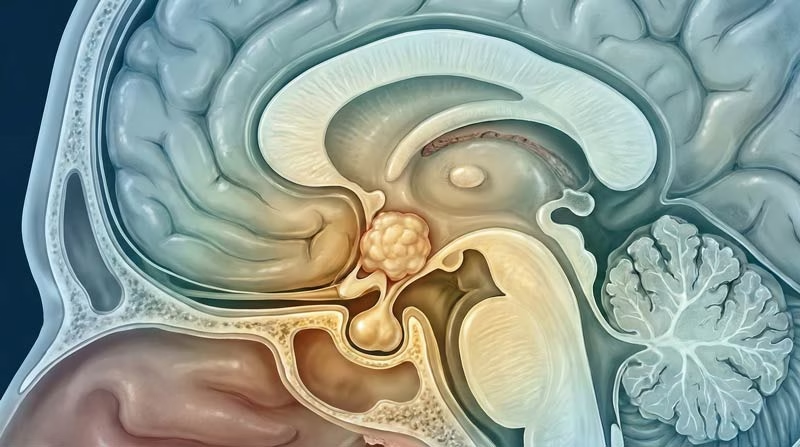
The most common tumors here are pituitary adenomas; craniopharyngiomas, meningiomas and trigeminal schwannomas also occur.
Back: near the brainstem
The back part, or posterior skull base, surrounds the brainstem and cerebellum. Specifically, a thick slope of bone called the clivus forms its center.
- Hearing loss or ringing in one ear
- Unsteady walking or dizziness
- Facial numbness or weakness
- Double vision and headaches at the back of the head
Typical tumors here include vestibular schwannomas (acoustic neuromas) and meningiomas, but also chordomas and chondrosarcomas.
Side: the ear and jugular foramen
The side of the skull base holds the ear and the jugular foramen. This opening carries the jugular vein and also the nerves for swallowing and voice.
- A pulsing sound in one ear, in time with the heartbeat
- Hoarseness or a weak voice
- Coughing or choking when swallowing
- A weak shoulder or tongue on one side
The most typical tumor here is the glomus jugulare tumor; schwannomas and meningiomas also occur.
Found by chance on a scan
Indeed, scans often show a small skull base tumor that causes no symptoms. In that case, surgery is rarely the first step.
Instead, doctors estimate the likely type and then repeat the MRI after a few months. If the tumor stays the same, many people then simply continue with regular scans.
Benign or malignant?
Most skull base tumors are benign, so they do not spread. According to MD Anderson, meningioma is the most common one. However, chordomas, chondrosarcomas and sinonasal cancers behave more aggressively.
How fast do symptoms appear?
- Slow tumors cause gradual changes over years.
- Cancers can cause symptoms within weeks.
- Bleeding into a pituitary tumor causes sudden headache and vision loss.
Questions at this stage
Can you feel a skull base tumor?
Usually not, because these tumors grow deep inside the head. However, a few extend into the neck and form a lump you can feel.
Do skull base tumors cause headaches?
Some do, especially when a tumor stretches the brain's covering or blocks fluid. Still, many never cause headaches, so vision, hearing and hormone changes matter more.
Are most skull base tumors benign?
Yes. For example, meningiomas, schwannomas and pituitary adenomas make up a large share, and all three are usually benign. Even so, a benign tumor can damage nerves as it grows.
When to See a Doctor
In fact, most of these symptoms have common causes, such as sinus or ear problems. Still, some patterns deserve a prompt check.
Book a doctor's visit if you notice:
- Hearing loss or ringing in one ear only
- A loss of smell that lasts without a cold
- Double vision or a drooping eyelid
- Nosebleeds from one side that keep returning
- Hormone changes, such as missed periods or new fatigue
Seek emergency care for:
- A sudden, severe headache with vision loss
- Sudden double vision or facial weakness
- Choking or trouble breathing when swallowing
- Confusion or unusual drowsiness
Generally, an MRI with contrast answers these questions. If it shows a tumor, a neurosurgeon with skull base experience can then explain the options. You can also compare these signs with other brain tumour symptoms.
Common Types of Skull Base Tumors
Dozens of tumor types can grow at the skull base. However, a handful cause most cases, and each follows its own pattern.
| Tumor | Where it grows | Behavior | Usual first step |
|---|---|---|---|
| Meningioma | Any region, from the brain's covering | Usually benign, slow | Surgery, radiosurgery or scans |
| Vestibular schwannoma (acoustic neuroma) | Hearing and balance nerve | Benign, slow | Scans, radiosurgery or surgery |
| Pituitary adenoma | Pituitary gland | Benign; may make hormones | Endoscopic surgery or tablets |
| Craniopharyngioma | Above the pituitary gland | Benign but sticks to nearby tissue | Surgery, often with radiotherapy |
| Paraganglioma (glomus jugulare) | Jugular foramen and middle ear | Usually benign, rich blood supply | Surgery, radiosurgery or scans |
| Chordoma and chondrosarcoma | Clivus and nearby bone | Malignant, slow, tends to return | Surgery, then high-dose radiotherapy |
| Sinonasal cancer | Nose and sinuses | Malignant | Surgery with radiotherapy |
| Metastasis | Any skull base bone | Spread from cancer elsewhere | Radiotherapy, surgery or drug treatment |
Three common types with their own guides
Meningioma grows from the brain's covering and is the most common skull base tumor. Doctors often watch small ones, while larger ones need surgery or radiosurgery. Also, read the full meningioma guide.
Craniopharyngioma sits above the pituitary gland and often affects vision, growth and hormones. Surgery must also protect the hypothalamus above it. See what craniopharyngioma is and how surgeons treat it.
Glomus jugulare tumors grow in the jugular foramen and often start with a pulsing sound in one ear. Their rich blood supply therefore shapes the plan. Read about glomus jugulare tumor treatment.
Is a skull base tumor cancer?
Usually not, since most skull base tumors are benign. However, chordomas, chondrosarcomas, sinonasal cancers and metastases are malignant, so they require more than one treatment.
Skull Base Tumor Treatment Options Compared
Treatment aims to remove or control the tumor while protecting the nerves around it. So, the right option depends on the tumor type, size, location and symptoms.
| Option | How it works | Best suited for | Recovery |
|---|---|---|---|
| Endoscopic endonasal surgery | Camera and tools through the nose; no skin cut | Midline tumors: pituitary, clivus, front skull base | A few days in hospital; nasal crusting for weeks |
| Open microsurgery (craniotomy) | Small skull opening; microscope and nerve monitoring | Tumors that spread sideways or wrap around nerves | About a week in hospital; weeks of rest at home |
| Stereotactic radiosurgery | Many focused beams in one to five sessions | Small tumors or tissue left after surgery | Usually home the same day |
| Fractionated or proton radiotherapy | Daily doses over several weeks | Larger tumors, chordoma and cancers | Daily visits; tiredness during treatment |
| Observation | MRI at set intervals | Small tumors without symptoms | No recovery time |
Endoscopic or open surgery?
The route follows the tumor, not the other way around. For example, midline tumors often suit the nose route. In contrast, tumors beside the carotid artery or far to the side need an open approach.
- Through the nose: pituitary tumors, many craniopharyngiomas, clival chordomas
- No visible scar and a shorter stay
- Main risk: a spinal fluid leak
- Through a skull opening: ear and jugular foramen tumors, large side extensions
- A wide view around nerves and vessels
- Main trade-off: a longer recovery
Some large tumors also need both routes, in one or two stages. Also, some hormone-making pituitary tumors, such as prolactinomas, shrink with tablets instead of surgery.
Can a skull base tumor be removed through the nose?
Often, yes. Endoscopic surgery reaches many midline tumors through the nostrils, without a cut on the face or scalp. However, tumors that spread far to the side still need an open approach.
Risks of Skull Base Surgery
Every skull base operation carries some risk, because the tumor sits among vital nerves and vessels. Therefore, the team plans a specific safeguard for each risk.
- Spinal fluid leak after endonasal surgery: a tissue flap seals the opening.
- New nerve weakness, such as double vision, facial weakness or swallowing trouble: nerve monitoring lowers this risk.
- Hormone changes after surgery near the pituitary gland: blood tests guide replacement.
- Infection, such as meningitis: antibiotics treat it.
- Bleeding or stroke: rare, from injury to a major vessel.
In practice, the surgeon may also leave a thin piece of tumor on a vital nerve. Then, radiosurgery can treat that piece later with much less risk. Overall, these safeguards follow the same principles as modern brain tumor surgery.
Your treatment path
From Diagnosis to Recovery
Treatment follows four steps. In turn, each step answers one question, from what the tumor is to what comes next.
STEP 01
Diagnosis: what is it, and what does it touch?
Diagnosis starts with a full neurological exam, including vision, hearing, smell and facial movement. Then, imaging shows the tumor and its neighbors.
- MRI with contrast shows the tumor, the nerves and the brain around it.
- CT shows the bone, such as erosion or thickening.
- CT or catheter angiography maps the vessels and the tumor's blood supply.
- Hearing, vision and hormone tests record function before treatment.
- A biopsy, often through the nose, confirms the type when scans leave doubt.
Is MRI or CT better for skull base tumors?
Doctors use both. MRI shows soft tissue and nerves in detail, while CT shows the bone of the skull base. Together, they guide the surgical route.
STEP 02
Planning: which route is safest?
First, a skull base team reviews each case together. It usually includes a neurosurgeon, an ear, nose and throat surgeon, a neuroradiologist, an endocrinologist and a radiation oncologist.
Next, the team chooses the route with the fewest nerves in the way. For very vascular tumors, a neuroradiologist may also block the feeding vessels a few days before surgery.
Why do some tumors need embolization before surgery?
Some tumors, such as paragangliomas, have a very rich blood supply. Blocking the feeding vessels first reduces bleeding, so the surgeon can work more safely.
STEP 03
Surgery: removal with nerve protection
The goal is maximal safe removal. The surgeon therefore removes as much tumor as the nerves and vessels allow.
- Neuronavigation tracks the instruments against the MRI in real time.
- Nerve monitoring warns the surgeon near facial, eye and swallowing nerves.
- The microscope or endoscope gives a magnified view.
- A tissue flap or graft rebuilds the skull base at the end.
Will I have a visible scar?
Endonasal surgery leaves no visible scar. In contrast, open surgery uses a cut behind the hairline or near the eyebrow, which usually fades well.
STEP 04
Recovery and follow-up
Recovery depends on the route and on any nerve that needs time to heal. In Prof. Albayrak's practice, open surgery usually means 1-2 days in intensive care, then 3-6 days on the ward.
- After endonasal surgery: no nose blowing, straining or heavy lifting for several weeks.
- After open surgery: most patients walk on the first day and shower by the third day.
- Rehabilitation: speech, swallowing or balance therapy when a nerve needs time.
- Follow-up MRI: at set intervals, often for years.
When can I fly home after skull base surgery?
Your surgeon decides after the post-operative checks. After endonasal surgery, the skull base repair needs time to seal, so some patients wait longer before flying.
Skull Base Tumor Surgery in Istanbul

Prof. Dr. Serdar Baki Albayrak
Neurosurgeon · Brain Tumor and Skull Base Surgery
Skull base surgery means reaching a tumor through narrow corridors between nerves and major vessels. Therefore, choosing the right route shapes both safety and recovery.
27+Years
5,000+Operations
64Countries
Figures from Prof. Albayrak's professional profile, September 2026.
Request an AppointmentFrequently Asked Questions
What is the survival rate for skull base tumors?
It depends mainly on the tumor type. Most skull base tumors are benign, so long-term control is common. Malignant types, such as chordoma, need combined treatment and closer follow-up.
How long is recovery after skull base surgery?
Most patients return to light activity within a few weeks. However, full recovery takes longer when a nerve needs time to heal.
Can a skull base tumor come back?
Some can. For example, chordomas and cancers carry a higher risk than benign tumors, so MRI follow-up continues for years.
Which doctor treats skull base tumors?
A neurosurgeon with skull base training leads treatment, often with an ear, nose and throat surgeon. In Istanbul, Prof. Dr. Serdar Baki Albayrak treats these tumors. He completed advanced surgical training at Harvard Medical School (Brigham and Women's Hospital) and a clinical fellowship in Helsinki.
Prof. Albayrak's Published Research
Peer-reviewed articles and book chapters written or co-written by Prof. Dr. Serdar Baki Albayrak on skull base and brain tumor surgery. Each link opens the record on PubMed or at the publisher. The full list of his 33 publications is on his profile.
Public health information
- National Cancer Institute. Chordoma.
- National Institute on Deafness and Other Communication Disorders. Vestibular Schwannoma (Acoustic Neuroma) and Neurofibromatosis.
This page gives general medical information and does not replace a consultation. Diagnosis and treatment decisions need an examination by your own doctor.
Plan Your Treatment in Istanbul
Request an appointment with Prof. Dr. Serdar Baki Albayrak. The clinical team then guides international patients through every step of the visit.
Request an Appointment