
Paid online consultations are available by appointment. For booking, please contact us via Whatsapp.

What Is Myelomeningocele?
Myelomeningocele is the most severe form of spina bifida. Part of the spinal cord and its nerves bulge into a sac on the back. Skin does not cover the sac, so the nerves stay exposed.
It forms in the first month of pregnancy, when the neural tube fails to close. Nerves below the gap then work poorly, which affects the legs, bladder and bowel. Generally, the higher the gap, the greater the effect.
Most babies also have a Chiari II malformation, and many develop hydrocephalus. However, with modern care, most children live into adulthood and go to school.
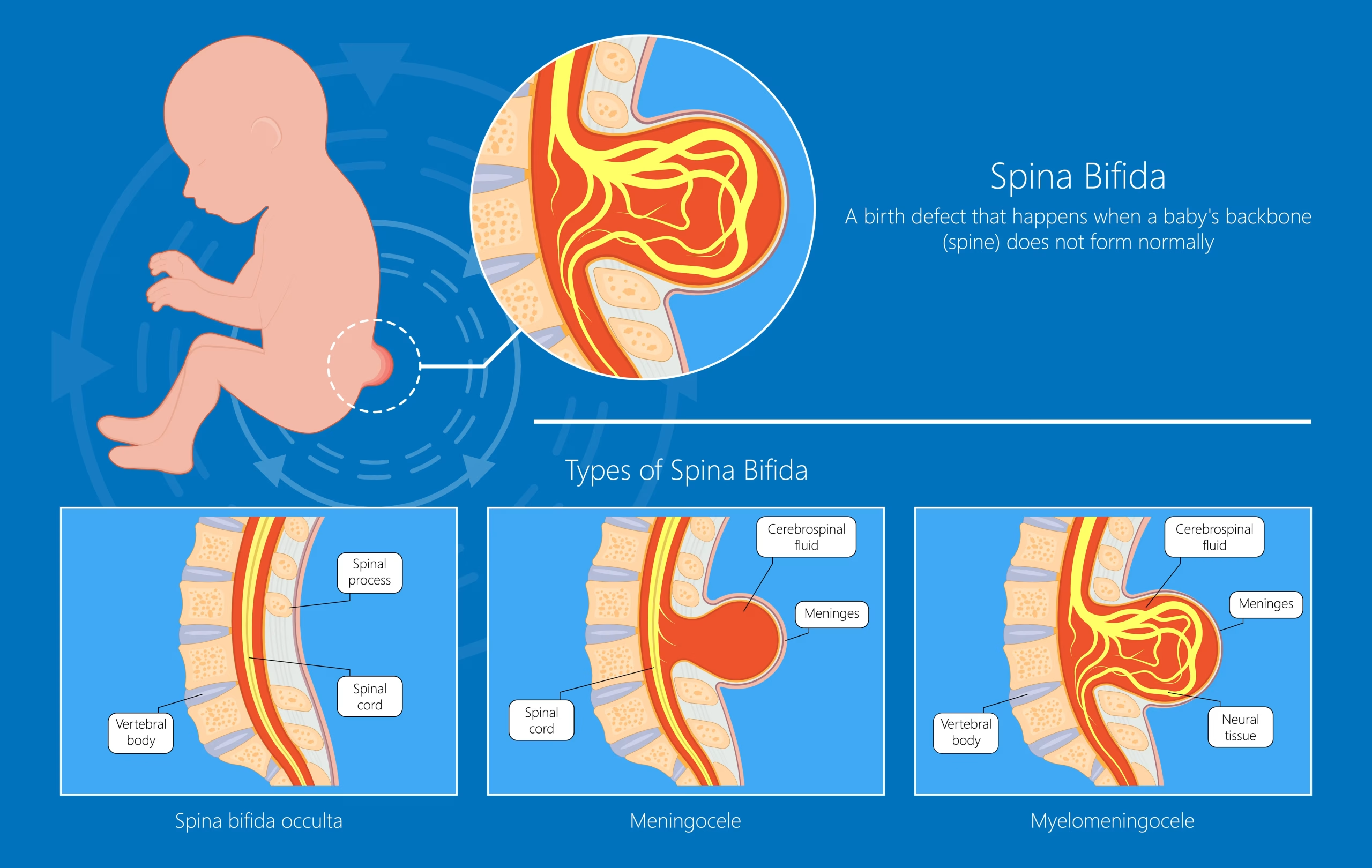
Myelomeningocele, meningocele or occulta: what is the difference?
In myelomeningocele, the sac holds nerve tissue, so some nerve damage is certain. In contrast, a meningocele holds only fluid and membranes. Spina bifida occulta is just a small gap in the bone, under normal skin.
Overall, myelomeningocele accounts for about 75% of spina bifida cases. Also, a flat open lesion without a sac, called a myelocele, needs the same urgent closure.
Where Are You Right Now?
First, choose the stage closest to yours. Then, each line opens the part of this guide that fits it.
- 01We just saw it on a pregnancy scanBlood test, ultrasound and fetal MRI
- 02We are choosing fetal or after-birth repairWhat the MOMS trial showed, and who qualifies
- 03Our baby was just born with an open sacThe first hours and the closure operation
- 04Our baby's head is growing fastHydrocephalus: shunt or ETV
- 05New problems after the repairChiari II, tethered cord and latex allergy
- 06We want to know about walking and the bladderOutlook by lesion level, school and adulthood
How Do Doctors Find Myelomeningocele Before Birth?
Most cases come to light in the second trimester. First, a blood test raises the question. Then, ultrasound and fetal MRI confirm it.
Maternal blood test (AFP)
Between weeks 16 and 18, a blood test measures alpha-fetoprotein (AFP). In fact, the level runs high in about 75-80% of pregnancies with spina bifida. So, a high result leads to a detailed scan.
Detailed ultrasound
The mid-pregnancy scan, around weeks 18-22, finds most cases. For example, it shows the typical "lemon" and "banana" head signs of the Chiari II malformation. In addition, it checks leg movement, the feet and the brain's fluid spaces.
Fetal MRI and genetic tests
Next, fetal MRI confirms the level of the lesion and the hindbrain herniation. Therefore, fetal surgery teams use it to decide who qualifies.
Amniocentesis checks the chromosomes, because other genetic conditions sometimes occur alongside. However, it remains optional, and your doctor explains its small risk first.
Questions at this stage
Can a scan miss myelomeningocele?
Rarely, yes. A very small lesion can stay hidden until birth. However, the detailed mid-pregnancy scan finds most cases.
Should we see a pediatric neurosurgeon during pregnancy?
Yes, ideally. A meeting with a pediatric neurosurgeon before birth explains the outlook, the delivery plan and both repairs.
Fetal Surgery or Repair After Birth?
Both repairs close the back and protect the exposed nerves. However, fetal repair happens before 26 weeks of pregnancy. In contrast, standard repair takes place within 48 hours of birth.
| MOMS trial result | Fetal repair (before 26 weeks) | Repair after birth |
|---|---|---|
| Shunt for hydrocephalus in the first year | About 4 in 10 babies | About 8 in 10 babies |
| Hindbrain herniation gone at 12 months | 36% | 4% |
| Walking without braces or crutches at 30 months | 42% | 21% |
| Average age at birth | 34.1 weeks | 37.3 weeks |
| Breathing problems from early birth | 21% | 6% |
| Effect on the mother | About 1 in 3 had a thin uterine scar at delivery; later births need a planned cesarean | No uterine scar from the repair |
So, fetal repair improves some outcomes, but it adds risks for mother and baby. Therefore, it takes place only at experienced fetal centers.
Who can have fetal surgery?
Also, fetal centers follow strict criteria. For example, the UCSF fetal team lists these:
- A lesion that starts between T1 and S1
- A Chiari II malformation on MRI
- Normal chromosomes and referral between 19 and 26 weeks
- A single baby, no short cervix and a body mass index of 35 or less
Open fetal surgery works through an opening in the uterus. In contrast, fetoscopic repair uses a few small ports, which may lower the risk to the uterus. However, fewer centers offer it.
Does fetal surgery cure spina bifida?
No. It can prevent further damage in the womb, but it cannot reverse damage that has already happened. So, many children still need a shunt or later surgery.
Hydrocephalus After Myelomeningocele: Shunt or ETV?
About 8 in 10 babies with myelomeningocele develop hydrocephalus, a build-up of fluid in the brain. It happens because the Chiari II malformation blocks the normal flow of spinal fluid.
Also, fluid can build up at birth or in the weeks after closure. So, the team measures head size often and repeats head ultrasounds.
Shunt
First, a thin tube with a valve drains fluid from the brain into the belly. It works for most babies, but it can block or become infected.
ETV with choroid plexus cauterization
In contrast, an endoscope opens a new path for fluid and reduces fluid production. As a result, it avoids a lifelong implant, but it suits only selected babies.
For more on both operations, see hydrocephalus in our pediatric neurosurgery guide.
Does every baby with myelomeningocele need a shunt?
No. For example, Great Ormond Street Hospital reports that about 60% of these children need a shunt. After fetal repair, the rate is lower still.
Chiari II, Tethered Cord and Other Problems Later On
Myelomeningocele itself does not grow, but related problems can appear months or years later. Therefore, children stay in regular follow-up.
Chiari II malformation
Almost every child with myelomeningocele has a Chiari II malformation. In it, the lower brain sits low, at the top of the neck. Many children never have symptoms from it.
However, watch for noisy breathing, breathing pauses, a weak cry or trouble swallowing. First, doctors check the shunt, because a blocked shunt can cause the same signs. Then, if symptoms continue, decompression surgery relieves the pressure.
Tethered cord and syrinx
Scar at the repair site can tether the spinal cord again, especially during growth spurts. In fact, the AANS estimates that 20-50% of these children need an untethering at some point.
Warning signs include new back pain, changes in walking, a worsening curve or a new bladder pattern. Also, a fluid cavity in the cord (syrinx) can cause similar signs. Our tethered cord surgery guide then explains the operation.
Latex allergy
Children with spina bifida have a much higher risk of latex allergy. So, hospitals use latex-free gloves and equipment from birth. Families also avoid latex at home and at school.
Can myelomeningocele cause problems later in life?
Yes. A tethered cord, shunt problems, a syrinx or scoliosis can appear years later, often during puberty. So, regular checks find them early.
When to Call the Doctor
Most days are routine. Still, some changes need a prompt call, and a few need emergency care.
Call your child's team if you notice:
- Late crawling or walking, or a change in walking
- New back pain, a worsening spinal curve or foot changes
- A new bladder pattern, frequent urine infections or constipation
- Red or broken skin on areas without feeling
Seek emergency care for:
- A bulging soft spot, vomiting, sleepiness or irritability
- Headache, vomiting or double vision in a child with a shunt
- Noisy breathing, breathing pauses or choking
- Fever with a stiff neck or a high-pitched cry
- Sudden weakness in the arms or legs
Walking, Bladder and Life With Myelomeningocele
The level of the lesion is the best guide to future walking. In contrast, bladder and bowel problems affect most children at every level.
| Lesion level | Muscles that usually work | Typical walking in childhood | Common aids |
|---|---|---|---|
| Thoracic | Upper body; little or no hip movement | Walking for daily use is rare; standing frames help | Wheelchair; hip-knee-ankle braces in early school years |
| High lumbar (L1-L3) | Hip flexors; some knee straightening at L3 | Short walks with braces and a walker or crutches | Wheelchair for longer distances |
| Low lumbar (L4-L5) | Knee straightening; some ankle movement | Many walk in the community | Ankle-foot braces; crutches or a wheelchair for long distances |
| Sacral | Most leg muscles | Often walk without braces | Foot orthotics for some |
However, walking often gets harder in the teens, as weight and height rise. So, many teenagers use a wheelchair for long distances and still walk at home.
Bladder and bowel care
Most children have a neurogenic bladder, so a urologist sees the baby soon after birth. Regular emptying with a thin tube, called catheterization, protects the kidneys. As a result, most children become dry with treatment.
Also, a bowel program with diet and medicines helps most children stay clean.
School, independence and adulthood
Most children have normal intelligence and attend school. Some find attention, planning or visual tasks harder, so early learning support helps.
Most people born with open spina bifida live into adulthood. Moreover, many go on to college and work.
Can a baby with myelomeningocele walk?
Many can, depending on the lesion level. For example, children with sacral or low lumbar lesions often walk, with or without braces. In contrast, higher lesions usually mean a wheelchair for most daily mobility.
How long do people with myelomeningocele live?
With early treatment and good kidney care, most people live into adulthood. Kidney problems from poor bladder drainage remain the main long-term risk, so bladder care matters for life.
What Causes Myelomeningocele, and Can You Prevent It?
Myelomeningocele comes from a mix of genetic and environmental factors. Importantly, nothing the parents did caused it. However, low folate before and in early pregnancy clearly raises the risk.
- Low folic acid before and in early pregnancy
- A previous pregnancy with a neural tube defect
- Certain anti-seizure medicines, such as valproic acid
- Poorly controlled diabetes, or obesity before pregnancy
- A high body temperature in early pregnancy, for example from a long fever or a sauna
Therefore, taking folic acid every day, starting before pregnancy, lowers the risk of neural tube defects. After an affected pregnancy, doctors prescribe a higher dose for the next one.
Is myelomeningocele hereditary?
Usually not. About 95% of affected babies have no family history. However, after one affected pregnancy, the chance of another rises to about 2-3%.
Your treatment path
From Pregnancy to Lifelong Care
Care follows four steps. Each step has one clear goal, so your family always knows what comes next.
STEP 01
Pregnancy: confirm and plan
After the diagnosis, a team of specialists meets the parents. Together, they review the scans, compare the repair options and plan the delivery.
Also, birth should take place in a hospital where the repair can happen quickly. As a result, the baby avoids a long transfer.
Is a cesarean delivery necessary?
After open fetal repair, yes, a planned cesarean protects the uterine scar. Without fetal repair, your obstetrician chooses the safest way to deliver.
STEP 02
Birth: protect the sac
First, right after birth, the team covers the sac with a sterile, moist dressing. Then, the baby lies on the tummy or side, and staff use latex-free equipment.
Next, the neurosurgeon checks leg movement, head size and the back. Meanwhile, a head ultrasound looks for hydrocephalus.
STEP 03
Repair: close the back within 48 hours
Under general anesthesia, the neurosurgeon frees the nerve tissue and places it back inside the spinal canal. Then, the surgeon closes the dura watertight and covers it with muscle and skin.
Also, for large defects, a plastic surgeon may help close the skin.
How long does myelomeningocele surgery take?
It usually takes a few hours. Specifically, the time depends on the size of the defect and the skin closure.
Can I hold and feed my baby after the operation?
Yes. The baby lies on the tummy most of the time. However, parents can usually feed and hold the baby.
STEP 04
Growing up: lifelong follow-up
Most babies stay in hospital for a week or more after the repair. Afterward, a team of neurosurgery, urology, orthopedics and physiotherapy follows the child.
Then, check-ups track head size, shunt function, the kidneys, walking, the spine and learning. Finally, in the late teens, care moves on to adult services.
When can international families fly home after the repair?
The surgeon decides once the wound heals and the baby feeds and grows well. For example, Great Ormond Street Hospital checks the wound and head size 10-14 days after surgery.
Myelomeningocele Care in Istanbul

Prof. Dr. Serdar Baki Albayrak
Neurosurgeon · Spina Bifida and Hydrocephalus Surgery
A newborn with myelomeningocele needs a fast, careful repair and a clear plan for the years ahead. Therefore, Prof. Albayrak works with the family from the first consultation through follow-up.
27+Years
5,000+Operations
64Countries
Figures from Prof. Albayrak's professional profile, September 2026.
Request an AppointmentFrequently Asked Questions
Is myelomeningocele the same as spina bifida?
Myelomeningocele is one type of spina bifida, and the most common open form. Spina bifida also includes meningocele and spina bifida occulta, as our spina bifida surgery guide shows.
Can myelomeningocele be cured?
No. Surgery closes the back and protects the remaining nerves, but it cannot restore damaged nerves. However, good care lets most children lead active lives.
What is the ICD-10 code for myelomeningocele?
In ICD-10, myelomeningocele falls under Q05, spina bifida. The fourth digit shows the level and any hydrocephalus. For example, Q05.2 means lumbar spina bifida with hydrocephalus.
Prof. Albayrak's Published Research
Peer-reviewed articles and book chapters written or co-written by Prof. Dr. Serdar Baki Albayrak on tethered spinal cord surgery in children. Each link opens the record on PubMed or at the publisher. The full list of his 33 publications is on his profile.
Public health information
- MedlinePlus (US National Library of Medicine). Myelomeningocele.
- Eunice Kennedy Shriver National Institute of Child Health and Human Development. Surgery on Fetus Reduces Complications of Spina Bifida (MOMS trial, 2011).
This page gives general medical information and does not replace a consultation. Diagnosis and treatment decisions need an examination by your own doctor.
Plan Your Child's Spina Bifida Care in Istanbul
Request an appointment with Prof. Dr. Serdar Baki Albayrak. The clinical team then guides your family through every step of the visit.
Request an Appointment