
Paid online consultations are available by appointment. For booking, please contact us via Whatsapp.

In short
What Is a Brain AVM?
A brain arteriovenous malformation (AVM) is a tangle of abnormal blood vessels. Inside it, arteries pour blood straight into veins, so there are no capillaries in between. An AVM is not a tumor or cancer, but it can cause bleeding.
Normally, capillaries slow blood down before it reaches the veins. In an AVM, however, high-pressure arterial blood hits thin-walled veins directly. Over time, these veins can stretch, and some eventually burst.
Brain AVMs affect fewer than 1 in 100 people, and most people carry them from birth. Yet symptoms usually appear in young adults, most often in their 20s to 40s.
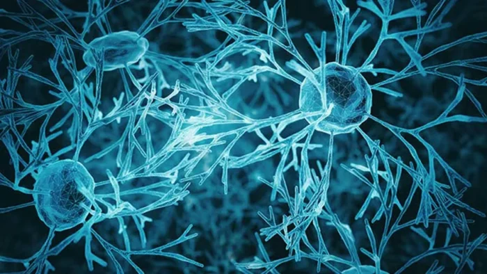
What are the parts of an AVM?
Every AVM has the same basic parts. Their size and position also decide how risky treatment will be.
- Feeding arteriesBring high-pressure blood into the tangle
- NidusThe tangle itself; its size drives the grade
- Draining veinsCarry blood out, to the surface or deep inside
- AneurysmsBulges on feeders or in the nidus raise the bleeding risk
- Nearby brainMay get too little blood, the so-called "steal" effect
- LocationAreas for movement, speech or vision count as "eloquent"
Which Symptom Brought You Here?
First, pick the change you or your family noticed. Then, each link explains how an AVM can cause it.
- 01A sudden headache or a brain bleedThe most common first sign, often with vomiting or drowsiness
- 02A seizureThe second most common first sign, often in younger adults
- 03Headaches that keep coming backSometimes in the same spot, sometimes migraine-like
- 04Weakness, numbness, vision or speech changesSlowly worsening problems from pressure or poor blood supply
- 05An AVM found by chanceNo symptoms, but a decision to make
Brain AVM Symptoms
Many AVMs cause no symptoms for years. When they do, bleeding and seizures are the two most common first signs.
The first sign often comes suddenly. About half of patients first learn about their AVM through a bleed. For others, a seizure or slowly worsening symptoms lead to the diagnosis.
Bleeding in the brain
An AVM bleed causes a sudden headache, often with vomiting, weakness, confusion or drowsiness. The exact signs depend on where the blood collects.
AVM bleeds tend to be smaller than aneurysm ruptures. However, each bleed carries about a 10-20% risk of death and a similar risk of lasting disability. After a first bleed, the yearly risk of another rises, especially during the first year.
Seizures
Seizures are the second most common first sign of an AVM. For example, they may start as jerking of one hand or the face, or spread into a full convulsion.
A first seizure in an adult always needs an MRI. Also, when seizures continue despite medicines, the plan borrows from epilepsy surgery. Removing the AVM often improves seizure control.
Recurring headaches
Headaches with an AVM vary widely. Some feel like migraine; others, however, keep returning in the same spot.
Yet a headache alone rarely points to an AVM. So doctors look for other signs, such as seizures or a whooshing sound in the head.
Weakness, numbness, vision or speech changes
An AVM can steal blood from nearby brain tissue or press on it. As a result, problems develop slowly over months:
- Weakness or numbness on one side of the body
- Loss of part of the visual field
- Trouble speaking or understanding speech
- Memory or concentration problems
- Clumsiness and dizziness, when the AVM sits in the cerebellum or brainstem
These slow changes can improve after treatment, especially when the team acts early.
An AVM found by chance
More AVMs now turn up on scans done for other reasons. In fact, the share of AVMs found before any bleed has nearly doubled in recent decades.
A silent AVM needs a calm decision rather than a rushed one. The ARUBA section below explains why.
Who gets brain AVMs?
Men and women develop AVMs equally, and AVMs occur in all ethnic groups. Most AVMs are sporadic; however, hereditary hemorrhagic telangiectasia (HHT) raises the chance of having one.
Can pregnancy affect an AVM?
Yes. Pregnancy raises blood volume and pressure, so some women notice new or worse symptoms. It may also raise the chance of a bleed, so planning ahead with your neurosurgeon helps.
Questions at this stage
Is a brain AVM hereditary?
Usually not. Most AVMs appear without any family history. However, people with HHT have a higher chance of AVMs, so doctors often screen them.
Can an AVM go away on its own?
Almost never. Spontaneous closure happens only rarely, so no one can plan around it. In fact, some AVMs slowly enlarge over the years.
Can an AVM cause memory problems or personality changes?
It can, mainly after a bleed or when the AVM steals blood from nearby brain areas. Treatment and rehabilitation can then help these changes improve.
When to See a Doctor
Most headaches have other causes. However, some signs need a prompt check, and a few need emergency care.
Book a doctor's visit if you notice:
- Headaches that keep returning in the same spot
- A whooshing sound in time with your pulse
- Slowly worsening weakness, numbness or vision loss
- A family history of AVM or HHT
- An AVM found on a scan, even without symptoms
Seek emergency care for:
- A sudden, severe headache
- A first seizure
- Sudden weakness, numbness or speech loss
- Confusion, fainting or unusual drowsiness
- Vomiting with a stiff neck
In an emergency, a CT scan shows a bleed within minutes. Next, an MRI and a catheter angiogram map the AVM itself. If the bleed came from a bulging artery instead, read about brain aneurysms.
Spetzler-Martin Grade: How Surgeons Rate an AVM
Surgeons grade every AVM before choosing a treatment. The Spetzler-Martin scale adds points for three features, so a higher total means a higher surgical risk.
- Size of the nidus: under 3 cm = 1 point, 3-6 cm = 2 points, over 6 cm = 3 points
- Eloquent location: areas for movement, sensation, speech or vision, plus the thalamus, hypothalamus and brainstem = 1 point
- Deep venous drainage: any drainage into the deep veins = 1 point
| Grade | What it usually means | Typical approach |
|---|---|---|
| Grade I-II (1-2 points) | Small, often near the surface and away from critical areas | Surgery often cures it with low risk; radiosurgery suits small, deep ones |
| Grade III (3 points) | A mixed group: small but deep, or large but superficial | A tailored plan, often combining embolization, surgery or radiosurgery |
| Grade IV-V (4-5 points) | Large, deep or inside critical brain | Surgery carries high risk; teams often observe unless bleeding repeats or deficits grow |
| Grade VI | Considered inoperable | Observation, with treatment only for specific problems |
Many teams also use the Lawton-Young supplementary scale. Younger age, a previous bleed and a compact nidus all lower the surgical risk, too.
What is a grade 3 AVM?
A Spetzler-Martin grade III AVM scores 3 points. However, it can be small and deep or large and superficial, so the risk differs widely. Therefore, teams tailor the plan for each grade III AVM.
Brain AVM Treatment: Surgery, Embolization, Radiosurgery or Observation?
Treatment aims to remove or close the whole nidus. A partly treated AVM can still bleed. So the team chooses one method, or a staged combination, for each AVM.
| Option | How it works | When teams choose it | Protection from bleeding | Main trade-off |
|---|---|---|---|---|
| Microsurgical removal | Through a craniotomy, the surgeon closes the feeding arteries, removes the nidus and divides the draining vein last | Grade I-II and selected grade III AVMs; AVMs with a large blood clot | Immediate, once an angiogram confirms complete removal | Open surgery; the risk rises with grade and location |
| Endovascular embolization | A catheter delivers a glue-like liquid that blocks feeders and parts of the nidus | Mostly before surgery or radiosurgery; alone only for some small AVMs | Only when the AVM closes completely | Risk of stroke or bleeding; partial embolization alone does not protect |
| Stereotactic radiosurgery | Focused radiation slowly thickens the vessel walls until the nidus closes | Small AVMs, usually under about 3 cm, deep inside or in critical areas | Delayed: closure takes 1-3 years, and the bleeding risk stays until then | Radiation changes in nearby brain; less effective in large AVMs |
| Observation (medical management) | Seizure medicines, blood pressure control and regular imaging | Many unruptured AVMs, especially high-grade ones or in older patients | None; the natural risk continues | A yearly bleeding risk for life |
After a bleed, teams may first remove a large blood clot and treat the AVM weeks later. Sometimes, however, the surgeon removes the clot and the AVM together.
Can a brain AVM be cured?
Yes, if the whole nidus disappears. Complete surgical removal cures the AVM at once, while radiosurgery can cure small AVMs over 1-3 years. Finally, an angiogram confirms the cure.
Treat or Watch an Unruptured AVM? What ARUBA Showed
For an AVM that never bled, no simple answer exists. ARUBA, the only randomized trial, favored watching, yet its limits keep the debate open.
ARUBA compared medical care alone with medical care plus surgery, embolization or radiosurgery. After about 33 months, 10.1% of the medical group had a stroke or died, versus 30.7% with treatment. Moreover, longer follow-up kept a similar gap.
However, follow-up covered only a few years of a lifelong risk. Also, most treated patients had embolization or radiosurgery rather than surgery.
For example, pooled data show that an unruptured AVM bleeds at about 2.2% a year. At that steady rate, the chance of a bleed over 20 years reaches about one in three.
In practice, many centers therefore watch unruptured AVMs of high grade. Meanwhile, they still offer surgery for small, low-grade AVMs in young patients.
- Your age and the years of risk ahead
- The grade and location of the AVM
- High-risk features: a deep location, only deep venous drainage or an aneurysm on a feeder
- Your own values about the risk of a procedure today versus a lifetime risk
AVM, Aneurysm, Cavernoma or Fistula: What Is the Difference?
These four vessel problems often get confused, yet they behave very differently. So the type decides both the risk and the treatment.
| Condition | What it is | Blood flow | Typical first sign | Main treatments |
|---|---|---|---|---|
| Brain AVM | A tangle of arteries and veins without capillaries | Fast, high pressure | Bleeding or a seizure in a young adult | Surgery, embolization, radiosurgery or observation |
| Brain aneurysm | A bulge on the wall of one artery | Arterial pressure inside a pouch | A sudden, severe headache if it ruptures | Clipping, coiling, flow diverter or observation |
| Cavernoma | A cluster of small, leaky blood-filled caverns | Very slow, low pressure | A seizure or a small bleed | Observation or surgery; radiosurgery only in select cases |
| Dural arteriovenous fistula | A direct artery-to-vein link in the brain's covering | Fast, into a vein or venous sinus | Pulsatile tinnitus, eye symptoms or a bleed | Embolization, surgical disconnection or radiosurgery |
In summary, an AVM stands out because it has a nidus inside the brain and high flow through it.
Your treatment path
From Diagnosis to Recovery
Treatment follows four steps. Each step then answers one question, from what the AVM looks like to how you recover.
STEP 01
Diagnosis: mapping the AVM
First, a CT or MRI shows the AVM and any bleeding. Then, a catheter angiogram (DSA) maps every feeding artery and draining vein, so it remains the gold standard.
- MRI shows the nidus, old bleeding and the brain around it.
- Functional MRI and tractography locate movement and speech areas near the AVM.
- Catheter angiography shows flow, venous drainage and any aneurysm on the feeders.
Can an MRI alone diagnose an AVM?
An MRI usually shows it clearly. However, treatment planning needs a catheter angiogram, because only it shows the flow pattern in detail.
STEP 02
Planning: choosing the safest route
Next, the team grades the AVM and weighs its natural risk against each treatment. For larger AVMs, the plan may combine steps, for example embolization before surgery.
This decision needs a team that can offer surgery, embolization and radiosurgery. Otherwise, the available tool, rather than the AVM, may drive the choice.
STEP 03
Treatment: removing or closing the nidus
During surgery, the surgeon works under the microscope with neuronavigation. Then, the order matters: feeding arteries close first, the nidus comes out next, and the draining vein goes last.
An angiogram during or after surgery confirms that no part of the nidus remains. For radiosurgery, in contrast, the team targets the nidus in a single session, often without an overnight stay.
Is AVM surgery dangerous?
It carries real risks, mainly bleeding and new weakness or speech problems. The risk depends mostly on the grade, so low-grade AVMs carry far less risk than high-grade ones.
STEP 04
Recovery and follow-up
In Prof. Albayrak's routine after planned brain surgery, patients spend 1-2 days in intensive care. Afterward, 3-6 days on the ward follow before discharge.
- Seizure medicines: many patients continue them for a while, and the neurologist decides when to stop.
- Surgery follow-up: an angiogram confirms the cure before you close the chapter.
- Radiosurgery follow-up: MRI scans continue until an angiogram shows complete closure.
How long is recovery after AVM surgery?
Many people return to light daily life within a few weeks after planned surgery. However, recovery after a large bleed takes months and often needs rehabilitation.
What happens after radiosurgery for an AVM?
The AVM closes slowly over 1-3 years. Until then, the bleeding risk remains, so you continue regular MRI scans. Finally, an angiogram confirms complete closure.
Arteriovenous Malformation Specialist in Istanbul
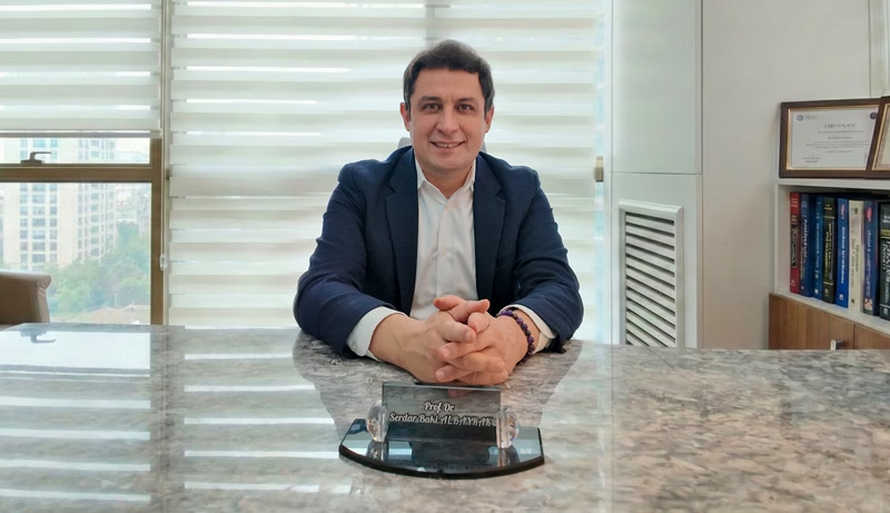
Prof. Dr. Serdar Baki Albayrak
Neurosurgeon · Cerebrovascular Surgery
AVM surgery rewards patience and order. The surgeon closes the feeders first and the draining vein last, so blood never gets trapped under pressure. Equally, deciding who should not have surgery matters as much as the operation.
His training includes advanced surgery at Harvard Medical School's Brigham and Women's Hospital. It also includes a clinical fellowship at the University of Helsinki.
27+Years
5,000+Operations
64Countries
Figures from Prof. Albayrak's professional profile, September 2026.
Request an AppointmentFrequently Asked Questions
How dangerous is a brain AVM?
On average, an unruptured AVM bleeds at about 2% a year. After a first bleed, however, the rate roughly doubles. Each bleed also carries a 10-20% risk of death.
What is the life expectancy with a brain AVM?
After complete removal or closure, most people can expect a normal lifespan. In contrast, without treatment the outlook depends on the yearly bleeding risk and on how each bleed affects the brain.
Can children have brain AVMs?
Yes, and bleeding often comes first. Since children face many years of risk, teams often favor treatment. They also follow children longer, because AVMs can recur after surgery at a young age; see pediatric neurosurgery.
Which doctor treats brain AVMs?
Generally, a cerebrovascular neurosurgeon leads care, together with an interventional neuroradiologist and a radiosurgery team. Prof. Dr. Serdar Baki Albayrak is a neurosurgeon in Istanbul whose practice includes brain AVM surgery.
Prof. Albayrak's Published Research
Peer-reviewed articles and book chapters written or co-written by Prof. Dr. Serdar Baki Albayrak on cerebrovascular microsurgery. Each link opens the record on PubMed or at the publisher. The full list of his 33 publications is on his profile.
- Microsurgical management of pineal region lesions: personal experience with 119 patients
- Microneurosurgical management of middle cerebral artery bifurcation aneurysms
- Microneurosurgical management of proximal middle cerebral artery aneurysms
Public health information
- National Institute of Neurological Disorders and Stroke. Arteriovenous Malformations (AVMs).
This page gives general medical information and does not replace a consultation. Diagnosis and treatment decisions need an examination by your own doctor.
Plan Your AVM Treatment in Istanbul
Request an appointment with Prof. Dr. Serdar Baki Albayrak. The clinical team then guides international patients through every step of the visit.
Request an Appointment