
Paid online consultations are available by appointment. For booking, please contact us via Whatsapp.

In short
What Is Craniopharyngioma?
A craniopharyngioma is a rare, slow-growing tumor that forms near the pituitary gland, usually just above it. It is benign, so it does not spread to other organs. However, it can press on the nerves of sight, the pituitary gland and the hypothalamus.
In fact, most craniopharyngiomas grow from cells left over from the gland's development before birth. Typically, the tumor has a fluid-filled cyst and a solid part, and many contain small calcium deposits.
As a result of its position, even a small tumor can cause big problems. For example, it can narrow side vision, stop a child's growth or cause constant thirst. Overall, surgery is the main treatment, and radiotherapy often follows it.
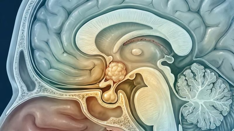
Which structures sit next to the tumor?
The tumor sits among six vital structures, so its symptoms depend on which one it presses on first.
- Pituitary glandMakes growth, thyroid, stress and sex hormones
- Pituitary stalkCarries the hormone that controls water balance
- HypothalamusSets hunger, thirst, sleep and body temperature
- Optic chiasmThe crossing point of the two optic nerves
- Third ventricleA fluid space the tumor can block
- Carotid arteriesLarge arteries on both sides of the tumor
Which Symptom Brought You Here?
First, pick the change you or your family noticed. Then, each link explains how a craniopharyngioma causes it.
- 01Blurred or narrowing visionBumping into things on one side, trouble reading or driving
- 02Constant thirst and frequent urinationDrinking large amounts, waking at night to pass urine
- 03Slow growth, late puberty or fatigueA child falling behind in height, missed periods, low energy
- 04Morning headaches and vomitingSigns of fluid building up inside the skull
- 05Weight gain, sleepiness or mood changeHunger that never settles, daytime sleepiness, memory problems
Craniopharyngioma Symptoms
Craniopharyngioma symptoms come from pressure on the nerves of sight, the hormone system and the brain's fluid pathways. The tumor grows slowly, so these symptoms often build up over months or years.
The first signs are easy to miss. For example, slow growth may look like a family trait, and tiredness may look like stress. As a result, the diagnosis often comes late.
Vision problems
The optic chiasm, where the two optic nerves cross, sits just above the pituitary gland. Therefore, a growing tumor often presses on it from below.
- Loss of the outer half of vision in both eyes (bitemporal hemianopia)
- Blurred vision or trouble reading
- Missing cars or people at the side when driving or walking
- Rarely, sudden vision loss when a cyst grows fast
However, children seldom complain about side vision. Instead, parents often notice clumsiness or falling school results first.
Constant thirst and urination
The hypothalamus makes antidiuretic hormone (ADH), and the pituitary stalk carries it. This hormone then tells the kidneys to save water. So, when the tumor damages this pathway, the body loses too much water.
- Large amounts of pale urine, day and night
- Strong thirst, often for cold water
- Bedwetting in a child who used to stay dry
Doctors call this central diabetes insipidus. Despite the name, however, it has nothing to do with sugar diabetes.
Slow growth and hormone problems
Pressure on the pituitary gland lowers one or more of its hormones. As a result, the symptoms depend on which hormone falls first.
- Growth hormone: a child grows much slower than classmates
- Sex hormones: late puberty, missed periods or low sex drive
- Thyroid hormone: tiredness, feeling cold, weight gain
- Cortisol: weakness, poor appetite, nausea and low blood pressure
Headaches and vomiting
A large tumor or cyst can block the fluid channels in the middle of the brain. Then, cerebrospinal fluid builds up, a condition doctors call hydrocephalus.
- Headaches that are worse in the morning
- Vomiting, sometimes without nausea
- Drowsiness or unsteady walking
Hydrocephalus therefore needs prompt care. In some cases, the surgeon drains the fluid first and removes the tumor a few days later.
Weight, sleep and behavior
The hypothalamus controls appetite, sleep, mood and body temperature. So, when the tumor presses on it, these daily rhythms can change.
- Fast weight gain from a hunger that never settles
- Daytime sleepiness and a disturbed sleep cycle
- Changes in memory, mood or behavior
- Trouble keeping a normal body temperature
Doctors call this weight gain hypothalamic obesity. It can also start after surgery, which explains why surgeons protect the hypothalamus so carefully.
Typical signs in children
- Slow growth or short height for age
- Headaches and vomiting from blocked fluid
- Vision loss that the child does not mention
- Late puberty in teenagers
Typical signs in adults
- Loss of side vision, often the first sign
- Low sex drive, missed periods or erection problems
- Tiredness and weight gain
- Memory or mood changes
Questions at this stage
What is usually the first sign of craniopharyngioma?
In children, headaches or slow growth often come first. In adults, a gradual loss of side vision is the most common first sign. Constant thirst can also appear at any age.
Can adults get craniopharyngioma?
Yes. The tumor has a second peak between about 50 and 74 years of age. Moreover, the papillary type occurs almost only in adults.
Can craniopharyngioma cause seizures?
Seizures are uncommon. However, they can occur with a large tumor, with a sharp drop in blood sodium or after surgery.
When to See a Doctor
In fact, most of these symptoms have simpler causes. Still, some patterns deserve a prompt check by a doctor.
Book a doctor's visit if you notice:
- A child who drops behind on the growth chart
- Side vision that seems to shrink in both eyes
- Constant thirst with large amounts of urine
- Headaches that keep coming back for weeks
Seek emergency care for:
- Sudden loss of vision
- A severe headache with vomiting or drowsiness
- Confusion or fainting
- Vomiting and weakness in someone who takes cortisol tablets
Generally, a brain MRI with contrast answers these questions quickly. If it shows a tumor, the next step is a consultation with a neurosurgeon who treats this region. Also, you can compare these signs with other brain tumour symptoms.
Types of Craniopharyngioma: Adamantinomatous vs Papillary
Craniopharyngiomas come in two types that look different under the microscope. The type also matters, because it predicts who gets the tumor and which medicines help.
| Feature | Adamantinomatous | Papillary |
|---|---|---|
| Who gets it | Mostly children; some adults | Almost only adults |
| Share of cases | About 9 in 10 tumors | About 1 in 10 tumors |
| On scans | Cysts, calcium and a fluid like machine oil | Mostly solid; calcium is rare |
| Border | Finger-like edges that stick to the brain | Usually a smoother border |
| Key gene change | CTNNB1 (beta-catenin) | BRAF V600E |
| Targeted medicine | None proven yet | BRAF and MEK inhibitor tablets |
In an NCI-funded trial, for example, these tablets shrank 15 of 16 papillary tumors.
Is craniopharyngioma cancer?
No. It is a benign tumor, so it does not spread to other parts of the body. Still, it can stick to vital structures and grow back, so it needs expert care.
Craniopharyngioma vs Pituitary Adenoma and Rathke Cleft Cyst
Several growths in this area can look alike at first. Examples include pituitary adenomas, Rathke cleft cysts and meningiomas. However, a few features usually tell them apart.
| Feature | Craniopharyngioma | Pituitary adenoma | Rathke cleft cyst |
|---|---|---|---|
| Where it starts | Above the gland, along the stalk | Inside the pituitary gland | Between the two lobes of the gland |
| On scans | Cyst plus a solid part, often calcium | Mostly solid, rarely calcium | A simple cyst without a solid part |
| Hormones | Low hormones; thirst from diabetes insipidus is more common | May make too much of one hormone | Usually normal |
| Usual plan | Surgery, sometimes radiotherapy | Medicine or surgery, depending on type | Scans, or drainage if it causes symptoms |
Craniopharyngiomas also behave more aggressively than pituitary adenomas, even though both are benign. For other tumors of this region, also see our guide to skull base tumors.
Craniopharyngioma Treatment Options Compared
Treatment relieves pressure and controls the tumor while protecting vision, hormones and the hypothalamus. Most patients need surgery first, and some also need radiotherapy.
| Option | How it works | Best suited for | Main trade-off |
|---|---|---|---|
| Endoscopic endonasal surgery | Camera and tools through the nose | Midline tumors below or behind the optic chiasm | No skin cut; small risk of fluid leak |
| Transcranial surgery | Small skull opening near the eyebrow or temple | Tumors spreading far sideways or upward | Wide side view; gentle brain retraction |
| Cyst drainage (Ommaya reservoir) | A tube drains the cyst into a scalp chamber | Large cysts, especially in young children | Quick relief; the solid part stays |
| Radiotherapy or proton therapy | Small daily doses over several weeks | Tumor left on purpose near the hypothalamus | Good control; late hormone effects possible |
| Stereotactic radiosurgery | One or a few focused high doses | Small leftovers away from the optic nerves | Short course; small targets only |
Can medicine help as well?
Yes, in selected cases. For example, doctors can place medicine into a cyst through the reservoir to slow its refilling. In addition, targeted tablets can shrink a papillary tumor with the BRAF change.
Overall, these methods fit into a wider plan of brain tumor treatment, built around each patient.
Can craniopharyngioma be treated without surgery?
Sometimes, especially when a papillary tumor carries the BRAF change. Otherwise, most patients need at least a limited operation to confirm the diagnosis and relieve pressure.
Why Protecting the Hypothalamus Shapes the Plan
Hypothalamic damage can cause lifelong obesity, fatigue and sleep problems, even after a clean scan. Therefore, the hypothalamus decides how far surgery should go.
First, the team checks on MRI how closely the tumor touches the hypothalamus. Many surgeons use a simple three-level scale for this, such as the Puget grade.
Prof. Albayrak also follows a clear rule. He never sacrifices the hypothalamus to remove the last piece of tumor.
When complete removal makes sense
- The tumor does not stick to the hypothalamus
- A clear plane separates the tumor from the brain
- The tumor sits mainly below the optic chiasm
When a partial removal is the safer plan
- The tumor grows into the wall of the hypothalamus
- Tumor tissue wraps around small vital vessels
- Radiotherapy can safely treat the part left behind
In many cases, this combined approach also controls the tumor well. At the same time, it lowers the risk of lasting hormone and hypothalamic problems.
Craniopharyngioma in Children
Craniopharyngioma is one of the most common tumors near the pituitary gland in childhood. Most children also have the adamantinomatous type, often with a large cyst.
In a child, treatment also protects growth, puberty and learning, because the child has decades of life ahead. For example, doctors may drain a large cyst first and delay radiotherapy. Then, proton therapy can spare more healthy brain later.
- Growth hormone and puberty care from a pediatric endocrinologist
- Early support for appetite and weight
- School and learning support after treatment
- Regular eye checks and MRI scans into adulthood
Families traveling to Istanbul can also read more about pediatric neurosurgery, from the first visit to follow-up care.
Your treatment path
From Diagnosis to Recovery
Treatment follows four steps. In turn, each step answers one clear question, from diagnosis to lifelong care.
STEP 01
Diagnosis: what exactly is it?
Diagnosis starts with a neurological exam and questions about growth, vision and thirst. Then, three groups of tests complete the picture.
- MRI with contrast shows the cyst, the solid part and the hypothalamus.
- CT shows calcium deposits, a typical clue for this tumor.
- Blood and urine tests check pituitary hormones, sodium and water balance.
- An eye exam with visual field testing measures side vision.
Is a biopsy needed before surgery?
Usually not. The scan pattern is typical, so the pathologist confirms the type from the tissue that surgery removes.
STEP 02
Planning: which route protects the most?
Before surgery, the team studies how the tumor relates to the optic chiasm, the stalk and the hypothalamus. This map then decides the route, the goal and the plan for any tissue left behind.
- Route: through the nose for most midline tumors, or through a small skull opening for tumors that spread to the sides
- Goal: complete removal when safe, or a planned partial removal
- Hormone safety: cortisol cover before and during surgery when levels are low
- Fluid pressure: drainage first when hydrocephalus is severe
Which surgical approach is best for craniopharyngioma?
No single approach suits every tumor. For example, the endoscopic route suits many midline tumors, while open surgery reaches tumors that spread to the sides. The best choice depends on the tumor's shape and the surgeon's experience with both.
STEP 03
Surgery: remove as much as is safe
The surgeon usually empties the cyst first, then separates the solid part from nerves and vessels. Throughout, the aim is safe removal, not removal at any cost.
- Neuronavigation shows the tumor's position in real time.
- The endoscope gives a close, angled view behind the optic chiasm.
- A flap of nasal lining seals the skull base after endonasal surgery.
- Microsurgery through a small opening reaches tumors high in the brain.
Does surgery through the nose leave a scar?
No. The surgeon works through the nostrils, so there is no visible cut. Afterward, many patients have a blocked nose and crusting for a few weeks.
STEP 04
Recovery and hormone care
Most patients stay in hospital for about 4 to 6 days. During this time, the team checks sodium, urine output and hormones several times a day.
Water balance can swing in three phases over two weeks. First, the body loses too much water; next, it may hold water; finally, some patients develop lasting diabetes insipidus.
- Tablets replace low cortisol and thyroid hormone.
- Desmopressin tablets or spray control diabetes insipidus.
- Growth and sex hormones follow later.
- MRI scans check the result early, then at regular intervals.
Why do doctors check sodium so often after surgery?
Surgery near the stalk can upset the water-balance hormone. As a result, sodium can change quickly, so fast adjustments matter.
When can I fly home after craniopharyngioma surgery?
Your surgeon decides after the first checks. In general, patients fly once sodium stays stable, the hormone doses work and the nose heals well.
Craniopharyngioma Surgery in Istanbul
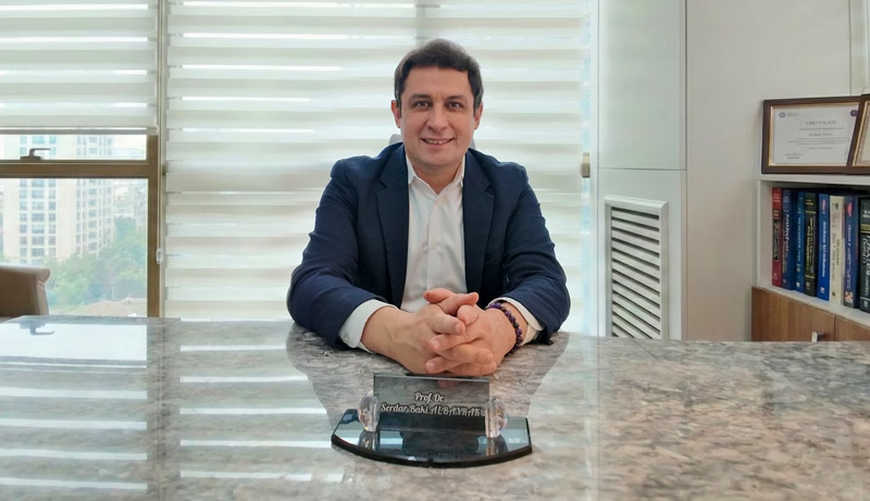
Prof. Dr. Serdar Baki Albayrak
Neurosurgeon · Brain Tumor and Skull Base Surgery
Craniopharyngioma surgery balances tumor removal against vision, hormones and the hypothalamus. Therefore, Prof. Albayrak plans the operation and the hormone care that follows as one treatment.
27+Years
5,000+Operations
64Countries
Figures from Prof. Albayrak's professional profile, September 2026.
Request an AppointmentFrequently Asked Questions
Can craniopharyngioma come back after surgery?
Yes, it can. Specifically, Cleveland Clinic reports regrowth in up to 17% after complete removal. After partial removal, the figure rises to 25-63%, so MRI follow-up continues for many years.
What is the life expectancy with craniopharyngioma?
Most people live many years after treatment. According to Cleveland Clinic, more than 90% of patients are alive five years after diagnosis. However, quality of life depends mostly on hormone and hypothalamic function.
Is craniopharyngioma hereditary?
No. It does not run in families, and nothing a parent or patient did caused it. It starts from cell changes during early development.
Which doctor treats craniopharyngioma?
A neurosurgeon with skull base experience leads treatment, together with an endocrinologist, an eye specialist and a radiation oncologist. In Istanbul, Prof. Dr. Serdar Baki Albayrak leads this care. He completed advanced surgical training at Harvard Medical School (Brigham and Women's Hospital).
Prof. Albayrak's Published Research
Peer-reviewed articles and book chapters written or co-written by Prof. Dr. Serdar Baki Albayrak on deep-seated brain tumors and image-guided surgery. Each link opens the record on PubMed or at the publisher. The full list of his 33 publications is on his profile.
- Microsurgical treatment of third ventricular colloid cysts by interhemispheric far lateral transcallosal approach: experience of 134 patients
- Intra-operative magnetic resonance imaging in neurosurgery
Public health information
- National Cancer Institute. Childhood Craniopharyngioma Treatment (PDQ), patient version.
- National Cancer Institute. Targeted drug combo may change care for rare brain tumor craniopharyngioma (2023).
This page gives general medical information and does not replace a consultation. Diagnosis and treatment decisions need an examination by your own doctor.
Plan Your Treatment in Istanbul
Request an appointment with Prof. Dr. Serdar Baki Albayrak. The clinical team then guides international patients and families through every step of the visit.
Request an Appointment