
Paid online consultations are available by appointment. For booking, please contact us via Whatsapp.

In short
What Is a Meningioma?
A meningioma is a tumor that grows from the meninges, the layers that cover the brain and spinal cord. Most are benign and generally grow slowly, over many years.
Overall, meningiomas are the most common primary brain tumor. In fact, they make up over a third of primary brain and spine tumors. They also affect women about twice as often as men.
Because it grows from the covering, a meningioma usually pushes the brain aside. As a result, surgeons can often remove it completely. Many patients then need no further treatment.
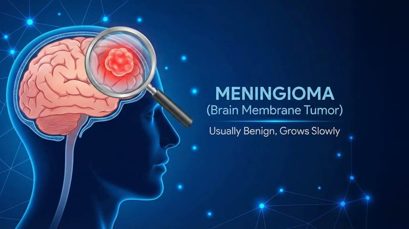
Where does a meningioma start?
The meninges have three layers, and the tumor starts in the middle one. It then anchors to the tough outer layer, which surgeons also remove.
- Dura materThe tough outer layer, where the tumor anchors
- ArachnoidThe thin middle layer, where meningioma cells start
- Pia materThe delicate inner layer on the brain surface
Which Symptom Brought You Here?
Meningioma symptoms depend mostly on where the tumor sits. First, pick what you noticed. Then, each link explains the likely cause.
- 01No symptoms: a scan found it by chanceCommon with small tumors seen on an MRI
- 02Headaches or a first seizureMorning headaches, or jerking of one hand or the face
- 03Vision, smell or hearing changesBlurred vision, a bulging eye, lost smell, one-sided hearing loss
- 04Weakness, memory or personality changeA clumsy leg, forgetfulness, apathy or poor judgment
- 05Unsteadiness, facial numbness or swallowing troubleSigns of a tumor near the brainstem
Meningioma Symptoms
Many small meningiomas cause no symptoms at all. When symptoms appear, they build up slowly, so people often adapt without noticing.
Found by chance
Many meningiomas turn up on scans for other problems, such as dizziness. In fact, autopsy studies find silent meningiomas in 1 to 2 percent of people. Therefore, the usual first step is a repeat MRI, not an operation.
Headaches and seizures
Meningioma headaches tend to be worse in the morning and grow more frequent over time. However, most headaches have harmless causes.
Seizures, in contrast, are a common first sign of tumors on the brain's surface. For example, a seizure may begin with jerking of one hand or the face.
Vision, smell and hearing
Tumors near the optic nerves blur vision or narrow the field of view. A sphenoid wing tumor can also push one eye forward. In addition, tumors beside the brainstem cause hearing loss or ringing in one ear.
Loss of smell often goes unnoticed, so olfactory groove tumors can grow large first.
Weakness, memory and personality
Parasagittal and falx meningiomas grow beside the brain's midline, next to the leg motor area. As a result, one leg may feel weak or clumsy first.
Large frontal tumors, in contrast, change behavior. Families often notice apathy or poor judgment first, as our frontal lobe tumors guide explains.
Balance, face and swallowing
Skull base meningiomas near the brainstem grow in a space crowded with cranial nerves. Therefore, they can cause facial numbness, unsteady walking, hoarseness or trouble swallowing. Our guide to skull base tumors also covers their surgery.
Meningioma symptoms by location
| Location | Typical symptoms | Surgical note |
|---|---|---|
| Convexity (outer brain surface) | Headaches, seizures, weakness | Usually easy to reach; complete removal is often possible |
| Parasagittal and falx (midline) | Leg weakness, seizures, personality change | Growth into the midline vein can limit removal |
| Sphenoid wing (ridge behind the eye) | Vision loss, bulging eye, double vision | May wrap around the carotid artery and optic nerve |
| Olfactory groove, tuberculum sellae (above the nose) | Loss of smell, personality change, vision loss | Some suit an endoscopic route through the nose |
| Posterior fossa (beside the brainstem) | Hearing loss, facial numbness, unsteadiness | Surgery and radiosurgery often work together |
| Spinal (mostly chest level) | Back pain, numbness, leg weakness | See spinal cord tumors |
Questions at this stage
How fast does a meningioma grow?
Generally, grade 1 meningiomas grow slowly, over years rather than months. Some stop growing altogether, especially calcified tumors in older adults. In contrast, grade 2 and 3 tumors can grow noticeably within months.
Can a meningioma cause dizziness or vertigo?
Sometimes: tumors near the brainstem, the cerebellum or the hearing nerve can upset balance. However, most dizziness has other causes. Your doctor therefore checks whether the tumor's position explains it.
When to See a Doctor
Most headaches, dizzy spells and vision changes have other causes. However, some patterns deserve a prompt neurological check and an MRI.
Book a doctor's visit if you notice:
- Headaches that change pattern or get worse over weeks
- Slowly blurring vision, or a loss of smell without a cold
- Hearing loss or ringing in one ear
- New clumsiness, forgetfulness or a change in personality
Seek emergency care for:
- A first seizure
- A sudden, severe headache
- Sudden weakness, numbness or speech loss
- Rapid loss of vision, confusion or unusual drowsiness
If you already have a meningioma, then report any new symptom to your doctor. Also, compare these signs with other brain tumour symptoms.
Meningioma Grades: WHO Grade 1, 2 and 3
The WHO grade shows how aggressive the tumor cells look under the microscope. As a result, it guides treatment and follow-up.
| Grade | How common | What the pathologist sees | Usual treatment |
|---|---|---|---|
| Grade 1 (benign) | About 80% | Slowly dividing cells, no brain invasion | Monitoring, surgery or radiosurgery; complete removal often ends treatment |
| Grade 2 (atypical) | About 18% | More dividing cells, brain invasion, or clear cell and chordoid subtypes | Surgery, often followed by radiotherapy after partial removal |
| Grade 3 (anaplastic) | Under 2% | Very rapid cell division, cancer-like cells, or TERT promoter or CDKN2A/B gene changes | Surgery followed by radiotherapy; drugs mainly in clinical trials |
Since 2021, gene tests can also decide the grade. For example, a TERT promoter mutation alone makes a tumor grade 3. Grade 2 and 3 tumors come back more often, so they need closer MRI follow-up.
Is there a grade 4 meningioma?
No. The WHO system has only three grades for meningioma. Grade 3 is the most aggressive form, but it is rare.
What Causes a Meningioma?
Most meningiomas start from random gene changes, with no single cause. However, several risk factors raise the chance:
- Previous radiation to the head, especially in childhood, the clearest known cause
- Neurofibromatosis type 2 (NF2-related schwannomatosis), which often causes several meningiomas
- Hormones: long-term use of some progestogen medicines, including the contraceptive injection, raises the risk
- Older age, with a sharp rise after 65
- Obesity, which several studies link to a higher risk
Is meningioma hereditary? Usually not, although people with NF2 inherit a strong tendency.
Watch, Operate or Radiosurgery: How Doctors Decide
Meningioma treatment ranges from MRI checks to surgery or focused radiation. The right choice then depends on symptoms, growth, location, grade and general health.
| Option | Best suited for | What it involves | Main trade-off |
|---|---|---|---|
| Active monitoring | Small tumors without symptoms, especially in older adults | An MRI after 3 to 6 months, then yearly if stable | No treatment risk, but regular scans |
| Microsurgery (craniotomy) | Tumors that cause symptoms, grow or press on the brain | Removing the tumor and its attachment | Often curative, yet it carries surgical risks |
| Stereotactic radiosurgery | Tumors under about 3 cm, deep skull base tumors, leftover tumor | One focused radiation session, usually outpatient | Controls most grade 1 tumors, which stay in place |
| Fractionated radiotherapy | Larger tumors near the optic nerves; grade 2 or 3 tumors after surgery | Daily sessions over several weeks | Gentler on nerves, but many visits |
Does a small meningioma need surgery?
Not always. In fact, size alone rarely decides, because many small tumors never cause harm. Therefore, doctors weigh five points together:
- Symptoms: seizures, vision loss or weakness favor treatment
- Growth: steady growth on serial MRI scans also favors treatment
- Location: tumors near the optic nerve or brainstem leave less room to wait
- Swelling: edema around the tumor signals brain irritation
- Age and health: a calcified, stable tumor in an older adult may never need surgery
In Prof. Albayrak's practice, surgery is the first choice for most tumors that cause symptoms. Generally, an operation takes 3 to 4 hours. In contrast, complex skull base surgery can last 12 hours or more.
Can a meningioma be treated without surgery?
Yes. Monitoring suits many small tumors, and radiosurgery or radiotherapy can also control others. However, no tablet or chemotherapy reliably shrinks a benign meningioma today.
Simpson Grade and the Risk of Recurrence
The Simpson grade describes how completely the surgeon removed the tumor and its attachment. Generally, the more complete the removal, the lower the chance of regrowth.
| Simpson grade | What the surgeon removed | Recurrence in Simpson's original series |
|---|---|---|
| Grade I | The whole tumor, its dural attachment and any abnormal bone | About 9% |
| Grade II | The whole tumor, with the attachment burned (coagulated), not cut out | About 16-19% |
| Grade III | The whole tumor, but not an attachment inside a large vein or bone | About 29% |
| Grade IV | Part of the tumor, leaving a piece behind | About 39-44% |
| Grade V | Only a biopsy or pressure relief | Not reported; regrowth expected |
Simpson published these figures in 1957, long before MRI. Since then, modern series have shown smaller gaps between grades I, II and III. Yet partial removal still carries a clearly higher risk.
Near cranial nerves, the brainstem or a major vein, removing every cell can cost function. In such cases, Prof. Albayrak may leave a thin layer and then treat it with radiosurgery.
Can a meningioma come back after surgery?
Yes, sometimes years later, so MRI checks continue long term. If it returns, the options include a second operation, radiosurgery or radiotherapy.
Your treatment path
From Diagnosis to Recovery
Meningioma care follows four steps. Each step then answers one question, from the first scan to long-term follow-up.
STEP 01
Diagnosis: what does the scan show?
An MRI with contrast is the key test. It shows a sharply outlined tumor on the dura, often with a thin "dural tail".
- CT scan shows calcium in the tumor and thickening of the nearby skull bone.
- MR venography shows whether the tumor narrows or blocks a large vein.
- Eye and hearing tests record a baseline when the tumor lies near those nerves.
Unlike most brain tumors, a meningioma rarely needs a biopsy first. The pathologist then confirms the diagnosis and grade after surgery. However, a dural brain metastasis occasionally looks similar.
STEP 02
Planning: the safest route
Before surgery, the team builds a 3D map of the tumor, veins and nerves. Then, neuronavigation uses this map to plan the smallest safe incision and skull opening.
Some large tumors have a rich blood supply. For these, a radiologist can first block the feeding arteries. As a result, this step, called embolization, can reduce bleeding.
STEP 03
Surgery: removing the tumor and its attachment
Under general anesthesia, Prof. Albayrak opens a small skull window and works under the microscope. First, he cuts the tumor's blood supply at its attachment. Next, he hollows out the tumor and lifts it away from the brain.
Finally, he removes the attachment and repairs the covering.
- Neuromonitoring tracks nerve and motor function near the skull base.
- Endoscopic surgery through the nose suits selected midline skull base tumors.
- Closure: a patch repairs the dura, and mini titanium plates fix the bone back in place.
What are the risks of meningioma surgery?
Risks include bleeding, infection, seizures, a spinal fluid leak and new weakness or nerve problems. Generally, convexity tumors carry a low risk. In contrast, skull base tumors carry more, because nerves and vessels crowd that area.
STEP 04
Recovery and follow-up
Most patients spend one night in intensive care. Usually, they stand up on the first day and can shower on the third. Most then go home after 3 to 5 days.
Next, the pathology result shapes the plan:
- Grade 1, complete removal: MRI follow-up only
- Grade 1, partial removal: monitoring or radiosurgery
- Grade 2 or 3: often radiotherapy, then closer MRI checks
Typically, the first MRI comes about three months after surgery. Afterward, scans follow yearly for several years.
How long is recovery after meningioma surgery?
Hospital stay is short, but full recovery takes longer. Even so, many patients feel close to normal within a few weeks. However, tiredness can last a few months, especially after long skull base operations.
Meningioma Surgery in Istanbul
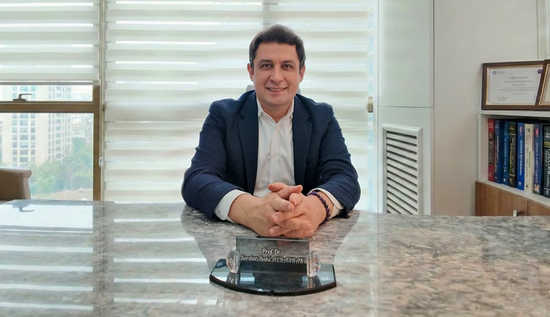
Prof. Dr. Serdar Baki Albayrak
Neurosurgeon · Brain Tumor and Skull Base Surgery
Removing the tumor's attachment lowers the risk of regrowth, and gentle technique also protects nearby nerves. When surgery is not the best first step, Prof. Albayrak also discusses monitoring or radiosurgery.
27+Years
5,000+Operations
64Countries
Figures from Prof. Albayrak's professional profile, September 2026.
Request an AppointmentFrequently Asked Questions
Is a meningioma cancer?
Usually not: about 8 in 10 are benign grade 1 tumors. In fact, fewer than 2 in 100 are malignant. Even so, a benign tumor can still harm the brain as it grows.
Can a meningioma be cured?
Often, yes. Complete removal of a grade 1 tumor with its attachment cures many patients. Where that is unsafe, radiosurgery keeps most remaining tumors under control.
What is the survival rate for meningioma?
It depends mostly on the grade and the extent of removal. For example, most people with a grade 1 meningioma return to normal life after treatment. For grades 2 and 3, however, the NCI reports a five-year relative survival of 63.5 percent.
Which doctor treats a meningioma?
A neurosurgeon experienced in brain tumor and skull base surgery usually leads care. Prof. Dr. Serdar Baki Albayrak treats patients with meningioma in Istanbul.
Prof. Albayrak's Published Research
Peer-reviewed articles and book chapters written or co-written by Prof. Dr. Serdar Baki Albayrak on meningiomas. Each link opens the record on PubMed or at the publisher. The full list of his 33 publications is on his profile.
- The Origin of Meningiomas
- The double immunostaining of CD133 and Ki-67 favours a significant co-localization pattern in fibroblastic subtype of meningiomas
- Postoperative cerebral abscess formation caused by Toxocara canis in a meningioma cavity
Public health information
- National Cancer Institute. Meningioma: Diagnosis and Treatment.
- National Cancer Institute. Adult Central Nervous System Tumors Treatment (PDQ), patient version.
This page gives general medical information and does not replace a consultation. Diagnosis and treatment decisions need an examination by your own doctor.
Plan Your Meningioma Treatment in Istanbul
Request an appointment with Prof. Dr. Serdar Baki Albayrak. The clinical team then guides international patients through every step of the visit.
Request an Appointment