
Paid online consultations are available by appointment. For booking, please contact us via Whatsapp.

What Is an Astrocytoma?
An astrocytoma is a tumor that grows from astrocytes. These star-like cells support the nerve cells of the brain and spinal cord. Astrocytomas belong to the glioma family, the most common tumors of brain tissue itself.
Some astrocytomas grow slowly for years, while others grow within months. So the exact type, the grade and the gene markers guide every treatment decision. Our overview of brain tumor treatment also covers other types.
Since 2021, the WHO system grades these tumors from 1 to 4 and adds gene tests. In particular, IDH status now separates astrocytoma from glioblastoma.
At a Glance
- What it isA glioma that grows from astrocytes, the support cells of the brain
- GradesWHO grade 1 to 4; gene tests such as IDH refine the grade
- First signsOften a first seizure; also headaches or slow changes in mood or strength
- DiagnosisMRI first, then tissue from surgery or a biopsy, with gene tests
- Main treatmentRemoval of as much tumor as is safe; then radiotherapy, chemotherapy or scans
- Follow-upRegular MRI scans for many years, more often at first
Where Are You Right Now?
First, pick the line closest to your situation. It then takes you to the matching part of this guide.
- 01A first seizure, headaches or a change in personalitySymptoms that can point to an astrocytoma
- 02An MRI shows a possible gliomaThe tests that confirm the type and grade
- 03A report that says pilocytic astrocytomaGrade 1, common in children and young adults
- 04A report that says IDH-mutant, grade 2 or 3What these grades mean for your treatment
- 05Grade 4, or the word glioblastomaHow grade 4 astrocytoma differs from glioblastoma
- 06Planning surgery from abroadLength of stay, flights and care back home
Astrocytoma Grades and Types
The WHO grade shows how fast a tumor tends to grow, from 1 (slowest) to 4 (fastest). Since 2021, gene markers also decide the grade, alongside the microscope. So the full name on your report matters, not only the number.
| Tumor type | WHO grade | How it behaves | Usual first steps |
|---|---|---|---|
| Pilocytic astrocytoma | 1 | Slow growth, often a clear border and a cyst | Surgery; often only MRI checks after full removal |
| Astrocytoma, IDH-mutant | 2 | Slow growth, but spreads into nearby brain | Surgery, then scans, a targeted pill, or radiotherapy and chemotherapy |
| Astrocytoma, IDH-mutant | 3 | Faster growth and more active cells | Surgery, then radiotherapy and chemotherapy |
| Astrocytoma, IDH-mutant | 4 | Fast growth; dead tissue or a CDKN2A/B gene loss | Surgery, then radiotherapy and chemotherapy |
| Glioblastoma, IDH-wildtype | 4 | A separate tumor type; fast growth, mostly in older adults | Surgery, then radiotherapy with chemotherapy |
Pilocytic Astrocytoma (Grade 1)
Pilocytic astrocytoma grows slowly and often has a clear edge. It also often holds a fluid-filled cyst and mostly affects children and young adults. Typical sites, for example, include the cerebellum, the optic nerves and the brainstem.
If the whole tumor comes out, NCI notes that close checks often replace further treatment. Radiotherapy may follow when some tumor remains. For the back of the brain, see cerebellar tumor surgery; for children, see pediatric neurosurgery.
Astrocytoma, IDH-Mutant: Grades 2 and 3
This group mostly affects younger and middle-aged adults. Its cells carry a change in the IDH1 or IDH2 gene. Most also lose the ATRX gene.
A grade 2 tumor grows slowly but spreads into nearby brain without a sharp edge. Over the years, however, some tumors turn into a higher grade, as NCI notes.
A grade 3 tumor, in contrast, shows more active, faster-dividing cells. Treatment then starts sooner, and scans come more often.
Grade 4 Astrocytoma and Glioblastoma
An IDH-mutant astrocytoma counts as grade 4 when it shows certain signs. These include dead tissue, new blood vessels or a loss of the CDKN2A/B gene. It often affects younger adults and tends to behave less aggressively than glioblastoma.
Glioblastoma, IDH-wildtype, in contrast, has no IDH change and now counts as a separate tumor. It mostly affects older adults and also grows fast. Our guide to brain cancer and glioblastoma covers it in depth.
Is astrocytoma cancer?
Mostly, yes. The NHS counts gliomas of grade 2 to 4 as cancer. Pilocytic astrocytoma (grade 1), however, usually behaves like a benign tumor.
Can an astrocytoma turn into glioblastoma?
An IDH-mutant astrocytoma can progress to a higher grade over years. Even at grade 4, however, doctors keep the name astrocytoma, IDH-mutant. Glioblastoma, IDH-wildtype, develops as a different disease from the start.
Astrocytoma Symptoms
Symptoms depend on where the tumor sits and how fast it grows. A slow tumor may cause no symptoms for years. Fast-growing tumors, in contrast, cause changes within weeks or months.
Seizures
A first seizure in an adult often leads to the diagnosis, especially with slower-growing tumors. It can range from a full convulsion to a brief twitch or a blank spell.
Seizure medicine usually controls them well. Removing the tumor can also reduce seizures.
Headaches, nausea and drowsiness
A growing tumor and the swelling around it raise the pressure inside the skull. So headaches often feel worse in the morning or ease after vomiting, as NCI describes.
Coughing or straining can also make the pain worse. Unusual sleepiness, too, calls for a prompt check.
Personality, memory and thinking
Family members often notice the change first: less drive, irritability or poor judgment. Memory and concentration may also slip.
Such signs typically come from the frontal or temporal lobe. Our guide to frontal lobe tumors explains them in detail.
Weakness, speech and vision
For example, weakness or numbness on one side can develop slowly. Some people struggle to find words or to follow speech. Others lose part of their field of vision.
Balance, swallowing and double vision
A tumor in the cerebellum, for instance, causes clumsiness and an unsteady walk. In the brainstem, double vision, swallowing trouble or facial weakness can appear. See our guide to brainstem tumors for that area.
Rarely, an astrocytoma grows in the spinal cord instead. Then back pain, weakness or numbness in the limbs lead; see spinal cord tumors.
Signs in children
Children often show morning headaches with vomiting, clumsiness or trouble walking. In babies, the head may grow too fast. School work can also slip.
Questions at this stage
What are the early signs of an astrocytoma?
Often a first seizure, especially with a low-grade tumor. Subtle changes in mood, memory or word finding may come first, too. Many slow tumors, however, cause no early signs at all.
When to See a Doctor
Most astrocytoma symptoms develop over weeks or months, so you usually have time to book a visit. Some signs, however, call for emergency care.
Book a doctor's visit if you notice:
- A first seizure, even if it stopped by itself
- Headaches that feel new or get worse each week
- Slow changes in personality, memory or speech
- Weakness or numbness on one side
- Blurred or double vision that does not go away
Seek emergency care for:
- A seizure that lasts over 5 minutes, or repeated seizures
- A sudden, severe headache
- Sudden weakness, numbness or trouble speaking
- Growing drowsiness or confusion
- Repeated vomiting with a headache
These signs can mean rising pressure in the skull or bleeding. So go to the nearest hospital first, rather than waiting for a visit abroad.
How Doctors Diagnose an Astrocytoma
An MRI scan with contrast dye generally shows the tumor best. Yet only tissue under the microscope, with gene tests, confirms the exact type and grade.
First, the neurosurgeon checks your strength, reflexes, balance, vision and speech. Then, depending on the case, these tests follow:
- MRI with contrast: size, edges, swelling and nearby structures
- MR spectroscopy and perfusion: hints of a higher grade inside the tumor
- Functional MRI and tractography: speech and movement areas and their pathways
- CT scan: bleeding, calcium or a quick check in an emergency
- PET scan: later on, telling regrowth apart from radiation changes
A scan alone can mislead, however. Prof. Albayrak co-authored a 2011 report on exactly this. In it, a lung cancer metastasis looked just like a cystic astrocytoma.
Pathology and Gene Markers
Tissue comes from surgery or, when removal carries too much risk, from a needle biopsy. A neuropathologist then names the tumor and its grade. Gene tests also follow on the same tissue, as NICE recommends.
| Marker | What it shows | Why it matters |
|---|---|---|
| IDH1 or IDH2 mutation | An astrocytoma or oligodendroglioma, not glioblastoma | Usually a slower course; a targeted pill for some grade 2 tumors |
| ATRX loss | Typical of IDH-mutant astrocytoma | Supports the diagnosis |
| 1p/19q codeletion | Present in oligodendroglioma, absent in astrocytoma | Changes the name and the chemotherapy plan |
| CDKN2A/B deletion | Loss of a gene that brakes cell growth | Raises an IDH-mutant astrocytoma to grade 4 |
| MGMT methylation | A DNA repair gene switched off | Predicts a better response to temozolomide |
| BRAF change | Typical of pilocytic astrocytoma | Opens the door to targeted drugs for some tumors |
What does IDH-mutant mean on my pathology report?
It means the tumor cells carry a change in the IDH1 or IDH2 gene. This change sets astrocytoma apart from glioblastoma, IDH-wildtype. It also usually points to a slower course.
What is the difference between astrocytoma and oligodendroglioma?
Both belong to the IDH-mutant gliomas. An oligodendroglioma, however, has also lost parts of chromosomes 1 and 19, the 1p/19q codeletion. An astrocytoma lacks it, so the two tumors get different chemotherapy plans.
Astrocytoma Treatment by Grade
Treatment depends on the type, grade, gene markers, location, and your age and health. First, surgery removes as much tumor as is safe. Then a tumor board plans what follows.
At this board, Prof. Albayrak reviews each case with oncology, radiotherapy and neurology colleagues.
| Tumor | After surgery | Points to weigh |
|---|---|---|
| Pilocytic, grade 1 | Usually MRI checks only; radiotherapy if tumor remains | Often curable when fully removed |
| IDH-mutant, grade 2 | MRI checks, a targeted pill, or radiotherapy with chemotherapy | Your age and any tumor left on MRI guide the choice |
| IDH-mutant, grade 3 | Radiotherapy, then chemotherapy | Treatment usually starts within weeks |
| IDH-mutant, grade 4 | Radiotherapy and chemotherapy | Closer MRI checks |
Radiotherapy
Radiotherapy aims precise beams at the tumor area and spares healthy brain as far as possible. Sessions then run on weekdays for several weeks. Modern methods include intensity-modulated radiotherapy (IMRT) and, for some tumors, proton therapy.
For low-grade tumors, doctors sometimes delay radiotherapy to protect memory and thinking. In children, they also weigh its effect on growth and development, NCI notes.
Chemotherapy and Targeted Pills
Temozolomide, taken as capsules, forms the most common chemotherapy for astrocytoma. For grade 3 astrocytoma, NICE recommends radiotherapy followed by up to 12 cycles of it. Another option, PCV, combines three drugs.
In 2024, the FDA approved vorasidenib, a daily pill, for grade 2 IDH-mutant astrocytoma after surgery. It blocks the faulty IDH enzyme and so can delay further treatment. Your oncologist then decides whether it suits you.
Does every astrocytoma need radiotherapy?
No. A fully removed pilocytic astrocytoma usually needs only MRI checks. NICE also suggests regular checks alone for people around 40 or younger whose IDH-mutant tumor came out completely.
Astrocytoma Surgery: Maximal Safe Resection
Surgery has three aims. It provides tissue for the diagnosis and relieves pressure on the brain. It also removes as much tumor as is safe.
With gliomas, research links a more complete removal to longer survival. Protecting speech, movement and vision, however, comes first.
The usual operation, a craniotomy, opens a small window in the skull under general anesthesia. Afterward, the bone goes back in place.
Tools That Help Protect Brain Function
Prof. Albayrak uses these methods, chosen for each tumor:
- Microsurgery: work under a high-power microscope, millimeter by millimeter
- Neuronavigation: a 3D map from your MRI that guides the route, much like GPS
- Fluorescence (5-ALA): a drink before surgery that makes high-grade tumor tissue glow under blue light
- Neuromonitoring: live checks of movement and speech pathways during removal
- Awake craniotomy: you wake during mapping, so the team can test speech and movement
Awake surgery mainly suits tumors near speech areas, such as many insular gliomas. The brain itself feels no pain, and the team stays with you throughout.
His 2004 review in Acta Neurochirurgica also describes intraoperative MRI. Such scans during surgery help especially in low-grade gliomas.
When a Biopsy Comes First
Sometimes an astrocytoma lies too deep or spreads too widely for safe removal. A stereotactic biopsy then takes a small sample through a tiny hole in the skull. That sample still allows the full gene tests.
Can an astrocytoma be completely removed?
A pilocytic astrocytoma often can, especially in the cerebellum. Diffuse astrocytomas, however, spread into nearby brain, so some tumor cells usually stay. Treatment after surgery then targets those cells.
Risks of Astrocytoma Surgery
Every brain operation carries real risks. Most serious problems stay uncommon, but you should know them before you decide.
Possible problems include:
- New weakness, numbness or speech trouble, often temporary
- Seizures in the first days
- Swelling of the brain, usually eased with steroid medicine
- Bleeding in the brain
- Infection of the wound or of the brain lining
- A spinal fluid leak from the wound
- Blood clots in the legs or lungs
Your own risk depends on the tumor's size and position, and on your health. So ask which risks matter most in your case.
Can surgery affect my speech or movement?
It can, especially near speech or movement areas. Many such problems improve over weeks with rehabilitation. Mapping and awake surgery also aim to keep this risk low.
Recovery and Follow-Up After Astrocytoma Surgery
Recovery usually starts with one or two days in intensive care, then several days on the ward. Most people walk with help on the first day. The final plan, however, waits for the pathology and gene results.
A Typical Timeline
| When | What usually happens | What to watch |
|---|---|---|
| Days 1-3 | First MRI within 72 hours; first short walks | Headache, drowsiness, wound swelling |
| Week 1 | Discharge, often with seizure and steroid medicine | New weakness or confusion |
| Weeks 2-3 | Pathology and gene results; tumor board plan | Tiredness |
| Weeks 3-6 | Radiotherapy or chemotherapy, if needed, often at home | Tiredness; hair loss where the beams pass |
| 3 months after radiotherapy | A new baseline MRI | Treatment changes can mimic growth |
How Often You Need MRI Scans
NICE gives a possible schedule by grade. Your own team may then adapt it:
- Pilocytic, fully removed: a scan at 12 months, then at growing intervals for 15 years
- IDH-mutant, grade 2: at 3 months, then every 6 months for 2 years; then yearly, and later every 1-2 years
- Grade 3 or 4: every 3-6 months for 2 years, every 6-12 months to year 4, then yearly to year 10
Treatment effects, however, can look like regrowth on early scans; our recurrent glioblastoma surgery guide explains how teams tell them apart. So advanced MRI or a PET scan sometimes helps tell them apart.
Living With an Astrocytoma
Tiredness often lasts for months, especially after radiotherapy. Physiotherapy, speech therapy and occupational therapy also help many people regain skills.
UK drivers must stop driving and tell the DVLA. After surgery for most IDH-mutant grade 2 or 3 astrocytomas, the break lasts at least a year. For grade 4 tumors, it lasts at least two years; elsewhere, ask about local rules.
Can an astrocytoma come back after surgery?
Yes, diffuse astrocytomas often regrow over time, because some cells stay behind. Pilocytic tumors, in contrast, seldom return after full removal. For this reason, regular MRI scans continue for years.
How long is recovery after astrocytoma surgery?
Most people go home within about a week and resume light tasks within a few weeks. Radiotherapy or chemotherapy, however, can add tiredness for months. Rehabilitation helps if surgery affects speech or strength.
Astrocytoma Prognosis: What Shapes the Outlook
No single number fits every astrocytoma. According to NCI, the outlook depends on the type, grade and gene changes. Location, leftover tumor and general health also matter.
Pilocytic astrocytoma often never returns after complete removal. IDH-mutant grade 2 tumors usually progress slowly, and many people live and work for many years. Grade 3 and 4 tumors need more intensive treatment, aiming at long-term control.
Is astrocytoma curable?
A pilocytic astrocytoma often is, once the surgeon takes it out completely. Diffuse astrocytomas, however, rarely go away for good. Even so, many people live with a controlled tumor for years.
What is the life expectancy with an astrocytoma?
It varies by grade, IDH status and age, so no figure fits everyone. IDH-mutant tumors generally do better than glioblastoma, IDH-wildtype. So ask your own team for an estimate, rather than relying on online averages.
Coming to Istanbul from the US, Canada, the UK or Europe
Astrocytoma care has two parts: surgery, then often months of radiotherapy, chemotherapy or scans. So many people have surgery in Istanbul and continue further treatment at home.
How Long to Stay in Istanbul
First, add up the consultation, any new scans, the operation and the hospital stay. Then rest near the hospital until your wound check. Overall, many plans come to about two weeks in Istanbul.
When You Can Fly Home
UK aviation guidance advises waiting about 7 days after brain surgery before flying. Air left inside the skull can expand at altitude. The US CDC also warns that flying soon after surgery raises the risk of blood clots.
Treatment and Follow-Up at Home
Before you leave, ask for the discharge summary, the full pathology report and copies of all scans. Your home team may also ask for the tissue slides. They can then repeat or extend the gene tests.
Next, book your oncology visit early, because radiotherapy often starts within weeks of surgery.
For insurance and Medicare, see brain surgery in Turkey: planning from the US. For NHS rules and travel insurance, see planning from the UK. From Canada, check with your provincial health plan and private insurer before you book.
Your treatment in Istanbul
From Your First Message to Flying Home
Your treatment in Istanbul follows four clear steps. Each step then answers one practical question.
STEP 01
Request an appointment
Use the form on this page, WhatsApp or call +90 532 308 97 72. A short message is enough: your diagnosis, your country and your travel dates.
You can write in English or Turkish, so use whichever feels easier. The team then agrees the consultation date with you.
STEP 02
Consultation and tests in Istanbul
First, Prof. Albayrak examines you at his clinic in Teşvikiye. Next, he goes through your MRI scans with you. If needed, newer scans can follow in Istanbul.
He then explains whether surgery or a biopsy suits you, and what may follow. Questions worth asking include:
- How much of the tumor can come out safely?
- Which functions lie close to the tumor?
- Which treatment will follow at home, and when?
What should I bring to my astrocytoma consultation?
First, bring your MRI scans on a disc or USB drive, with the written reports. Also bring any pathology report, a list of your medicines and notes on seizures. A relative who knows your symptoms helps, too.
STEP 03
Surgery and hospital stay
If you choose surgery, the operation takes place in a hospital in Istanbul. Prof. Albayrak then names the hospital at the consultation, along with the likely stay.
STEP 04
Recovery, fitness to fly and follow-up
After discharge, you rest at a nearby hotel, ideally with a companion. Then, before the flight, your surgeon checks the wound and confirms that you can fly.
Back home, get urgent help for any of these signs:
- A seizure, especially a long or repeated one
- A severe or worsening headache, or repeated vomiting
- New weakness, confusion or trouble speaking
- Fever, redness or fluid leaking from the wound
Can I have radiotherapy at home after surgery in Istanbul?
Yes, in most cases. Your home oncology team then plans it from the pathology report and the scans after surgery. So contact them before you travel, if you can.
Your Astrocytoma Surgeon in Istanbul
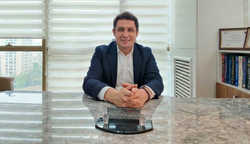
Prof. Dr. Serdar Baki Albayrak
Neurosurgeon · Full Professor of Neurosurgery
An astrocytoma often lies next to areas for speech or movement, so the margin for error stays small. For this reason, Prof. Albayrak combines microsurgery with navigation, fluorescence and nerve monitoring.
He completed his neurosurgery training at Istanbul University in 2004. He then took clinical fellowships at the University of Helsinki and Harvard Medical School. At Harvard, he trained at Brigham and Women's Hospital.
27+Years
5,000+Operations
64Countries
33Publications
Figures from Prof. Albayrak's professional profile, September 2026. See his full publication list.
Request an AppointmentFrequently Asked Questions
Is astrocytoma hereditary?
Usually not, and most astrocytomas have no known cause. A few inherited conditions, however, raise the risk, such as NF1, Li-Fraumeni syndrome and tuberous sclerosis. Past radiotherapy to the head also adds some risk.
Can an astrocytoma spread to other parts of the body?
Very rarely. An astrocytoma spreads within the brain and sometimes along the spinal fluid. Spread outside the brain and spine, however, almost never happens.
How much does astrocytoma surgery cost in Turkey?
The cost depends on the operation, the hospital stay and the tests you need. Gene tests, surgical tools such as fluorescence and your hotel stay also count. So ask for the cost of your own plan at the consultation, before you decide.
Prof. Albayrak's Published Research
None of Prof. Albayrak's papers focuses on astrocytoma treatment alone. The closest cover MRI during glioma surgery, an astrocytoma mimic and glioma research. Each link opens the record on PubMed; his profile lists all 33 publications.
- Intra-operative magnetic resonance imaging in neurosurgery
- Cerebral metastasis of small-cell lung carcinoma mimicking a supratentorial cystic astrocytoma
- The effects of tibolone on the human primary glioblastoma multiforme cell culture and the rat C6 glioma model
Public health information
- National Cancer Institute. Adult Central Nervous System Tumors Treatment (PDQ), patient version.
- National Cancer Institute. Childhood Glioma (Including Astrocytoma).
- National Institute for Health and Care Excellence. Brain tumours (primary) and brain metastases in over 16s (NG99): recommendations.
- US Food and Drug Administration. FDA approves vorasidenib for Grade 2 astrocytoma or oligodendroglioma with a susceptible IDH1 or IDH2 mutation.
- NHS. Brain tumours.
- UK Civil Aviation Authority. Surgical conditions: guidance for health professionals.
- US Centers for Disease Control and Prevention. Medical tourism.
- GOV.UK, Driver and Vehicle Licensing Agency. Neurological disorders: assessing fitness to drive.
This guide gives general medical information and does not replace a consultation. So diagnosis and treatment decisions need an examination by your own doctor.
Last updated: 1 October 2026. Website editor: Prof. Dr. Serdar Baki Albayrak's clinic, [email protected].
Plan Your Astrocytoma Consultation in Istanbul
Request an appointment with Prof. Dr. Serdar Baki Albayrak. The team then helps you plan the dates of your visit.
Request an Appointment