
Paid online consultations are available by appointment. For booking, please contact us via Whatsapp.

In short
What Is an Insular Glioma?
An insular glioma is a brain tumor that starts from glial cells in the insula, a lobe hidden deep inside the brain. Many grow slowly, but some are high grade.
Specifically, the insula sits under the frontal, parietal and temporal lobes, deep in the Sylvian fissure. In fact, it hosts up to a quarter of all low-grade gliomas.
Surgery here is demanding, because arteries that supply the movement pathways cross the tumor. However, modern mapping lets surgeons remove most tumors safely. As a result, maximal safe removal is now the usual first step.
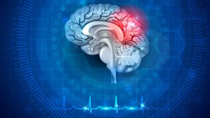
What does the insula do?
The insula links body signals with emotion, speech and awareness. Therefore, a tumor here can cause unusual, hard-to-describe symptoms.
- Speech planningThe dominant side, usually the left, helps plan speech
- Body awarenessHeartbeat, breathing and gut feelings reach awareness here
- Taste and smellThe central insula helps process both senses
- EmotionEmpathy, disgust and emotional reactions
- AttentionDecides which signals deserve attention
- Heart and blood pressureHelps adjust the body's automatic functions
Which Symptom Brought You Here?
Insular glioma symptoms often feel strange and hard to describe. First, pick what you noticed. Then, each link explains the cause.
- 01A seizure with an odd smell, taste or rising stomach feelingSometimes with throat tightness, drooling or a racing heart
- 02Trouble finding or saying wordsUsually with tumors on the left, speech-dominant side
- 03Weakness or numbness in the face or armPressure on the deep movement pathways
- 04Anxiety, emotional change or poor focusSubtle changes that family may notice first
- 05Already diagnosed and considering surgeryHow doctors decide, and what awake surgery involves
Insular Glioma Symptoms
Seizures are the first sign in most patients, especially adults under 40. Larger tumors can also cause headaches, nausea or drowsiness.
Seizures
Insular seizures often start with a sensation rather than jerking. For example, patients describe an odd smell or taste. Others also feel a rising stomach sensation or throat tightness.
Some also feel sudden fear, déjà vu or a racing heart. Consequently, doctors sometimes mistake these seizures for panic attacks or heart problems.
Speech and language
On the speech-dominant side, usually the left, the tumor can slow speech. It can also block word finding, especially near Broca's area.
Weakness and numbness
Large tumors can also press on the deep pathways that move the face and arm. As a result, one side may feel weak, numb or clumsy.
Emotion and attention
The insula shapes how we sense body signals and emotions. Therefore, some patients report anxiety, emotional flatness or trouble reading other people's feelings. These changes are often subtle, so families notice them first.
Left or right insula: does it matter?
Yes. Left-sided tumors more often affect speech, so surgeons usually plan awake mapping. In contrast, right-sided tumors tend to affect emotion and attention.
Can an MRI find it by chance?
Yes, sometimes during a scan for headaches or a minor injury. Even then, doctors usually recommend treatment, because gliomas tend to grow.
Questions at this stage
Can seizures stop after insular glioma surgery?
Often, yes. In fact, seizures improved in about 80 percent of patients in large surgical series. Some can then reduce or even stop their medicines.
Is an insular glioma cancer?
Gliomas grow into brain tissue, so doctors treat them as cancer. However, low-grade gliomas grow slowly, and many patients live for many years.
When to See a Doctor
Most odd smells or panic-like spells have other causes. However, repeated spells with the same pattern deserve a neurological check.
Book a doctor's visit if you notice:
- Repeated spells of an odd smell, taste or rising stomach feeling
- Brief episodes of déjà vu or fear without a trigger
- New trouble finding words
- Numbness or weakness on one side
Seek emergency care for:
- A first convulsive seizure
- A seizure lasting more than 5 minutes
- Sudden weakness or speech loss
- Severe headache with vomiting or drowsiness
Generally, an MRI with contrast answers the question. Also, compare these signs with other brain tumour symptoms. If seizures persist, read about epilepsy surgery too.
How Doctors Classify Insular Gliomas
Two systems matter before surgery: the tumor's molecular type and its position within the insula. Together, they also predict risk and outcome.
Tumor type and grade (WHO 2021)
| Tumor type | Key markers | WHO grade | What it means |
|---|---|---|---|
| Astrocytoma, IDH-mutant | IDH mutation, no 1p/19q codeletion | 2, 3 or 4 | Slower course than glioblastoma; the grade guides further treatment |
| Oligodendroglioma | IDH mutation plus 1p/19q codeletion | 2 or 3 | Often responds well to radiotherapy and chemotherapy |
| Glioblastoma, IDH-wildtype | No IDH mutation | 4 | The fastest-growing type; needs surgery, radiotherapy and chemotherapy |
Purely insular gliomas often carry an IDH mutation. In fact, one series found it in every purely insular tumor. In contrast, only 55 percent of tumors spreading into nearby limbic areas carried it.
Berger-Sanai zones: where in the insula?
First, two lines divide the insula: the Sylvian fissure and a vertical line through the foramen of Monro. The zone then predicts how much tumor surgeons can safely remove.
| Zone | Position | What covers it | Surgical note |
|---|---|---|---|
| Zone I | Front, upper part | Frontal operculum | Most often involved; usually allows the most complete removal |
| Zone II | Back, upper part | Motor and sensory strip for the face and arm | Often the hardest to remove completely |
| Zone III | Back, lower part | Temporal operculum, near hearing and language areas | Needs careful language mapping on the left |
| Zone IV | Front, lower part | Front of the temporal lobe | Usually reachable through the temporal lobe |
| Giant (I to IV) | All four zones | All overlying lobes | Staged or partial removal may be safer |
An older Yasargil system also separates purely insular tumors from those spreading into nearby lobes.
Which insular zone is the hardest to operate on?
Zone II often is, because the motor strip for the face and arm covers it. However, a mapped window through the cortex often lets surgeons reach it safely.
Insular Glioma Surgery: Who Needs It and How It Works
For most insular gliomas, surgery is the first step. It confirms the diagnosis, reveals the molecular type and often eases seizures too.
Decades ago, many surgeons avoided the insula and simply watched these tumors. Today, however, large series link more complete removal with longer survival. This also holds for both low-grade and high-grade gliomas.
| Approach | Route | Strengths | Limits |
|---|---|---|---|
| Transsylvian | Opens the Sylvian fissure between the frontal and temporal lobes | Spares the overlying cortex | Works directly among artery branches; narrower view |
| Transcortical | Small mapped windows in the overlying cortex | Wider exposure; safer for zone II in some series | Needs cortical mapping, often with the patient awake |
| Stereotactic biopsy | A needle through a small hole | Diagnosis when removal is unsafe | No tumor reduction |
| Laser ablation (LITT) | A laser probe heats the tumor from inside | Minimally invasive option for selected small tumors | Limited long-term data; nearby vessels restrict it |
Prof. Albayrak prefers awake surgery with brain mapping for tumors on the speech-dominant side. He also uses neuronavigation and continuous neuromonitoring in every insular operation.
Is insular glioma surgery dangerous?
It carries real risks, especially new weakness or speech problems. In fact, more than 1 in 5 patients have temporary deficits in modern series. Permanent deficits, however, stay below about 10 to 15 percent.
After Surgery: Markers and Further Treatment
Next, the pathology report guides everything that follows. Specifically, the IDH and 1p/19q results decide the type and the next step.
- Grade 2, IDH-mutant, well removed: MRI monitoring, or an IDH-inhibitor tablet (vorasidenib) to delay further treatment
- Grade 2 with higher risk, or grade 3: radiotherapy followed by chemotherapy
- Glioblastoma (grade 4): radiotherapy with temozolomide chemotherapy
Follow-up MRI scans then track the result, every few months at first.
What do IDH and 1p/19q mean?
IDH is a gene, and a mutation in it marks a slower-growing glioma. In addition, a 1p/19q codeletion means two chromosome arms are missing, which defines an oligodendroglioma.
Insular Glioma Survival Rate and Outlook
No single survival rate fits all insular gliomas. The outlook therefore depends on the molecular type, the grade and the extent of removal.
Linked to longer survival
- Seizures as the first symptom
- An IDH mutation
- Removal of 90 percent or more of the tumor
- Good general fitness before and after surgery
Linked to a harder course
- Higher grade, especially IDH-wildtype glioblastoma
- Spread into the putamen or nearby limbic areas
- Arteries wrapped by tumor, which limit removal
- Poor general fitness
For example, the NCI reports a five-year relative survival of 79.5 percent for oligodendroglioma. Glioblastoma, in contrast, remains far more serious, so treatment aims to extend good-quality life. Our guide to brain cancer and its surgery also covers it in detail.
What is the survival rate for insular glioma?
It depends on the molecular type and the extent of removal. For example, IDH-mutant low-grade tumors often allow many years of good-quality life. Your surgeon then explains your own outlook once the pathology result arrives.
Your treatment path
From Diagnosis to Recovery
Treatment follows four steps. Each step then answers one question, from the first scan to recovery.
STEP 01
Diagnosis: what kind of tumor is it?
First, an MRI with contrast shows the tumor, its edges and any faster-growing part.
- Perfusion MRI and MR spectroscopy estimate how aggressive the tumor is.
- EEG records seizure activity when seizures occur.
- Neuropsychological tests measure memory, language and attention as a baseline.
STEP 02
Planning: mapping language, pathways and arteries
Functional MRI shows which side controls language. Then, DTI tractography maps the nerve pathways around the tumor.
The team also studies the arteries, because the small lenticulostriate branches feed the deep motor pathways. Finally, it places the tumor in its zone and chooses the route.
STEP 03
Surgery: awake mapping and maximal safe removal
For left-sided tumors, Prof. Albayrak usually performs awake surgery. The patient sleeps during the opening, then wakes to talk and move during mapping.
- Direct electrical stimulation shows which areas still control speech and movement.
- Motor evoked potentials warn the team when a movement pathway is near.
- Neuronavigation guides each step, much like GPS.
Who is not suitable for awake surgery?
Awake mapping needs a calm patient who can follow simple tasks. However, severe speech loss, heavy anxiety or breathing problems may rule it out. In those cases, surgery then proceeds asleep with motor monitoring.
STEP 04
Recovery and further treatment
Most patients go home 3 to 4 days after surgery. Temporary speech or movement weakness often improves over weeks to months, and rehabilitation also helps.
Meanwhile, the pathology result sets the further plan, as described above.
How long do temporary speech problems last?
Most improve over weeks to months. However, recovery can take longer after surgery on the speech-dominant side, and speech therapy helps.
Insular Glioma Surgery in Istanbul
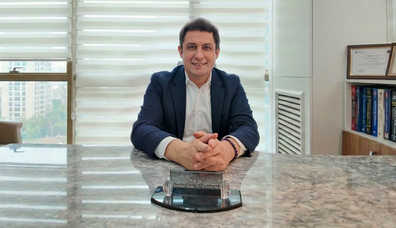
Prof. Dr. Serdar Baki Albayrak
Neurosurgeon · Brain Tumor and Skull Base Surgery
Insular surgery happens millimeters from the arteries that feed the movement pathways. Therefore, Prof. Albayrak combines awake mapping, neuronavigation and neuromonitoring to remove as much tumor as is safe.
27+Years
5,000+Operations
64Countries
Figures from Prof. Albayrak's professional profile, September 2026.
Request an AppointmentFrequently Asked Questions
Can an insular glioma be removed completely?
Sometimes, especially in zone I. More often, the aim is maximal safe removal, then other treatments. Overall, modern series remove a median of about 80 percent of the tumor.
Can an insular glioma come back?
Yes. Gliomas grow into the surrounding brain, so some cells usually remain. Regular MRI scans therefore catch regrowth early, when further treatment can still help.
Which doctor treats insular glioma?
A neurosurgeon experienced in awake glioma surgery leads care, with neuro-oncology and radiation teams too. Prof. Dr. Serdar Baki Albayrak treats insular gliomas in Istanbul.
For other tumor types, also see our brain tumor treatment guide. In addition, see our page on nearby frontal lobe tumors.
Prof. Albayrak's Published Research
Peer-reviewed articles and book chapters written or co-written by Prof. Dr. Serdar Baki Albayrak on glioma surgery and research. Each link opens the record on PubMed or at the publisher. The full list of his 33 publications is on his profile.
- Intra-operative magnetic resonance imaging in neurosurgery
- The effects of tibolone on the human primary glioblastoma multiforme cell culture and the rat C6 glioma model
Public health information
- National Cancer Institute. Oligodendroglioma and Other IDH-Mutated Tumors: Diagnosis and Treatment.
- U.S. Food and Drug Administration. FDA Approves Vorasidenib for Grade 2 Astrocytoma or Oligodendroglioma.
This page gives general medical information and does not replace a consultation. Diagnosis and treatment decisions need an examination by your own doctor.
Plan Your Insular Glioma Surgery in Istanbul
Request an appointment with Prof. Dr. Serdar Baki Albayrak. The clinical team then guides international patients through every step of the visit.
Request an Appointment