
Paid online consultations are available by appointment. For booking, please contact us via Whatsapp.

What Is Chordoma Surgery?
Chordoma surgery removes a slow-growing bone cancer from the skull base or the spine. The goal is to take the whole tumor out in the first operation, ideally in one piece with a rim of healthy tissue. High-dose radiotherapy then usually follows.
A chordoma grows from tiny leftovers of the notochord, the rod of tissue that shapes the spine before birth. So it can appear anywhere from the skull base to the tailbone. Most, however, sit at the two ends of this line.
However, chordoma rarely responds to standard chemotherapy, and radiation alone seldom controls it for long. For this reason, the first operation matters more here than in most cancers. A tumor that returns is much harder to treat the second time.
At a Glance
- What it isA rare, slow-growing bone cancer (sarcoma)
- Where it growsSacrum, skull base (clivus) and the spine between
- Typical signsDouble vision and headache, or tailbone pain
- Key testsMRI, CT, then a carefully planned biopsy
- Main treatmentSurgery, usually followed by high-dose radiotherapy
- Follow-upRegular MRI scans for many years
Where Are You in Your Decision?
Pick the line that sounds most like you. Each one then opens the matching part of this guide.
- 01A scan shows a mass and my doctor plans a biopsyWhy the biopsy route matters so much
- 02The biopsy confirmed chordoma and I need a planWhy the first operation shapes the outcome
- 03My chordoma is at the skull baseEndoscopic surgery through the nose and other routes
- 04My chordoma is in the sacrum or tailboneSacrectomy, nerve roots and bladder control
- 05Surgery, proton therapy or both?All treatment options side by side
- 06I am planning treatment from abroadLength of stay, flights and radiotherapy at home
Where Chordoma Grows and the Symptoms It Causes
In general, symptoms depend on where the tumor sits, and they build up slowly over months or years. According to MedlinePlus, about half of chordomas grow in the sacrum and about a third at the skull base.
Skull base (clival) chordoma
The clivus is a slope of bone just in front of the brainstem. So a tumor here first presses on the nerves that move the eyes. Double vision is often the first sign, together with a deep headache at the back of the head.
As it grows, trouble swallowing, a hoarse voice, facial numbness or a blocked nose can follow. Then, later on, pressure on the brainstem can affect balance and strength.
Sacral and tailbone chordoma
For months, a sacral chordoma may cause only low back or tailbone pain that gets worse when you sit. The tumor also grows forward into the pelvis, so nobody can feel it from outside for a long time.
Later, constipation, bladder changes or numbness in the legs and buttocks can appear. In fact, tailbone pain together with bowel or bladder change needs an MRI.
Neck, chest and lower back chordoma
In the mobile spine, too, chordoma usually causes steady local pain. If it presses on the spinal cord or a nerve root, numbness or weakness in the arms or legs follows.
The Three Locations Compared
| Feature | Skull base (clivus) | Sacrum and tailbone | Neck, chest, lower back |
|---|---|---|---|
| Share of cases | About one third | About half | The rest |
| Typical age | Younger adults; also children | Mostly over 50 | Middle age |
| First sign | Double vision, headache | Tailbone pain, constipation | Local pain, numbness |
| Nearby structures | Brainstem, cranial nerves, carotid artery | Bladder and bowel nerves, rectum | Spinal cord, vertebral artery |
| Removal in one piece | Rarely possible; maximal safe removal | Possible in many patients | Depends on the level |
Questions at this stage
Is chordoma cancer?
Yes. In fact, chordoma is a malignant bone tumor, a type of sarcoma. However, it grows slowly, and its main danger lies in local growth into nearby nerves, vessels and bone.
Can my tailbone pain be a chordoma?
Very rarely. Almost all tailbone pain comes from falls, long sitting or joint strain. Still, pain that lasts for months together with constipation, bladder change or numbness deserves an MRI.
When to See a Doctor About a Possible Chordoma
Generally, slow and steady symptoms deserve a specialist visit and an MRI. A few signs, however, mean that a nerve or the brainstem sits under pressure, and they call for emergency care.
Book a specialist visit if you notice:
- Double vision that slowly gets worse
- A deep headache at the back of the head that keeps coming back
- Tailbone pain for months, worse when you sit
- A bone mass on a scan done for another reason
Go to the nearest emergency department (911, 999 or 112) for:
- New or fast-rising trouble swallowing or speaking
- Weakness in the arms or legs that gets worse within days
- Loss of bladder or bowel control, or numbness between the legs
- Clear fluid dripping from the nose with fever after nasal surgery
Numbness between the legs with bladder change can signal pressure on the nerve bundle at the base of the spine. So never wait for an outpatient visit in that case.
How Doctors Confirm Chordoma, and Why the Biopsy Route Matters
Scans raise the suspicion, but only a tissue sample confirms chordoma. Above all, the surgeon who will operate should plan that biopsy.
| Test | What it shows | Why it matters |
|---|---|---|
| MRI with contrast | Tumor edges, nerves, brainstem or spinal cord | The base of the surgical plan |
| CT scan | Bone damage and calcium inside the tumor | Bone margins and the need for fixation |
| CT or MR angiography | Main arteries around the tumor | Carotid and basilar artery risk at the skull base |
| Whole-body scan | Spread to lungs, bone or liver | Sets the aim of treatment |
| Needle biopsy | Final diagnosis; brachyury staining | Tells chordoma apart from chondrosarcoma and metastasis |
The Biopsy Track
Specifically, chordoma cells can settle along the path of a biopsy needle. So the needle should pass through tissue that the surgeon will later remove with the tumor. For this reason, ask for the biopsy at the center that will treat you.
At the skull base, a biopsy through the nose can also serve as the start of the operation. In the sacrum, by contrast, the team avoids a biopsy through the rectum, because it can spread tumor cells into the bowel wall.
What is the difference between chordoma and chondrosarcoma?
Both can grow at the skull base, so they look alike on MRI. However, chordoma comes from notochord remnants, while chondrosarcoma comes from cartilage and usually sits slightly off the midline. Skull base chondrosarcoma often has a better outlook, and the pathologist tells them apart with brachyury staining.
Is chordoma hereditary?
Usually not. MedlinePlus notes that a few families carry extra copies of the TBXT gene, which makes the brachyury protein. So if two relatives have chordoma, your doctor may suggest genetic counselling.
Why the First Chordoma Surgery Shapes the Outcome
The strongest treatment for chordoma is removal in one piece with healthy margins in the first operation. NCI notes that outcomes vary with the tumor's location and with how much of it the surgeon removes.
In contrast, cutting a chordoma into pieces can spill cells into the wound, and new tumors can grow there. Also, scar tissue after a first operation hides the planes the surgeon needs. As a result, surgery for a recurrence is longer, riskier and less often complete.
Questions to Ask Any Chordoma Treatment Center
- Who plans the biopsy, and will its track come out with the tumor?
- Is removal in one piece possible in my case, or only maximal safe removal?
- Which nerves are at risk, and what would losing them mean for me?
- Who gives the radiotherapy, which type, and when does it start?
- How often will I have MRI scans, and for how many years?
Clear answers to these questions matter more than the name of a center. In addition, a team of neurosurgeon, radiation oncologist, radiologist and pathologist should make the plan together.
Chordoma Treatment Options Compared
Surgery comes first for most people, and radiotherapy then treats what remains. Chordoma resists ordinary doses, so radiation oncologists use techniques that focus a high dose on the tumor and spare the nerves around it.
| Option | How it works | Role in chordoma |
|---|---|---|
| Removal in one piece (en bloc) | Takes the tumor out with a rim of healthy tissue | The basis of long-term control, mostly in the sacrum |
| Maximal safe removal | Removes as much tumor as possible and spares vital structures | The usual approach at the skull base |
| Proton or carbon ion therapy | Leaves most of the dose in the tumor and little behind it | Often chosen near the brainstem and spinal cord |
| Intensity-modulated photon therapy | Shapes the X-ray beams to the tumor | A strong option when protons are not available |
| Stereotactic radiosurgery | A high dose to a small target in one to five sessions | Small leftovers or small recurrences |
| Targeted drugs and trials | Block the signals that drive tumor growth | Spread or inoperable disease, mostly in research |
Classic chordoma rarely responds to standard chemotherapy. On the other hand, a rare, fast-growing subtype, poorly differentiated chordoma, mostly affects children, and there chemotherapy has a clearer role.
Can proton therapy replace surgery for chordoma?
For most people, no. Proton therapy works best on the small amount of tumor left after surgery. So the team usually plans the widest safe removal first and high-dose radiotherapy after it.
Does chemotherapy work for chordoma?
Classic chordoma usually does not respond to it, so doctors seldom use it. Instead, for spread or inoperable disease, oncologists consider targeted drugs and clinical trials.
Clival Chordoma Surgery: Routes to the Skull Base
Particularly at the skull base, the tumor lies against the brainstem, cranial nerves and main arteries. So the aim is maximal safe removal, and the surgeon picks the route by the direction in which the tumor grows.
- Endoscopic surgery through the nose: the surgeon reaches the clivus through the nostrils, with no cut on the face; the same corridor serves pituitary tumor surgery
- Lateral skull base approaches: for tumors that extend sideways or behind the ear
- Combined or staged surgery: two routes, in one session or in two, for large tumors
Nerve Monitoring and Skull Base Repair
During the operation, the team checks the cranial nerves in real time with neuromonitoring. Then, after an endoscopic removal, the surgeon rebuilds the skull base in layers. Otherwise, brain fluid can leak into the nose.
Also, chordoma is only one of several tumors in this area. For the others, see our skull base tumor guide.
Can a surgeon remove a clival chordoma through the nose?
A surgeon can reach most midline clival chordomas through the nose with an endoscope. So you avoid a large cut on the face or head. If the tumor spreads sideways, however, the surgeon adds a lateral approach.
Will my double vision improve after surgery?
Often it does, once pressure comes off the nerve. Recovery can take weeks to months, and a nerve under long pressure may not recover fully. Prism glasses help in the meantime.
Sacral Chordoma Surgery: Sacrectomy and Nerve Roots
In the sacrum, the surgeon usually removes the tumor in one piece together with the affected part of the bone. This operation is a sacrectomy, and a general surgeon and plastic surgeon often join the team.
Here a key trade-off appears. Especially important, the sacral nerve roots run the bladder, the bowel and sexual function. So before surgery, the surgeon explains which roots the tumor involves and which ones the plan can keep.
What the Level of the Tumor Means
- Low tumors: when the third sacral roots stay intact on both sides, most people keep control of the bladder and bowel
- Higher tumors: losing more roots raises the chance of needing a catheter or a bowel routine
- Upper sacrum: removal weakens the link between spine and pelvis, so screws and rods rebuild it
For other tumors of the spine, see our spine and spinal cord tumors guide.
Will I lose bladder control after sacral chordoma surgery?
Mostly, that depends on which nerve roots the surgeon can keep. When the third sacral roots survive on both sides, most people keep control. If the tumor surrounds these roots, your surgeon discusses this risk with you in advance.
Can I walk after a sacrectomy?
Most people walk again, often with a frame or crutches at first. Physiotherapy starts in hospital, and the pace depends on how much bone and nerve the surgeon removed.
Chordoma Surgery Complications
Chordoma surgery is major surgery near vital structures, so it carries real risks. Therefore, your surgeon should explain each one for your tumor's exact location.
- Cranial nerve problems: double vision, swallowing or voice change after skull base surgery, often temporary
- Brain fluid leak: clear fluid from the nose after endoscopic surgery; it may need a second repair
- Artery injury: rare, but serious near the carotid or basilar artery
- Bladder, bowel and sexual function: after sacral surgery, linked to the nerve roots removed
- Wound healing: slower after sacral surgery and radiotherapy; plastic surgery flaps help
- Infection and blood clots: as with any long operation
What is the success rate of chordoma surgery?
No single figure fits everyone, because results depend on location, size and the margin the surgeon achieves. In general, removal with healthy margins in the first operation gives the longest control. So ask what to expect in your own case.
Chordoma Surgery Recovery Time
Generally, recovery after endoscopic skull base surgery is shorter than after a sacrectomy. Radiotherapy then adds several weeks to the plan.
| Stage | Skull base (endoscopic) | Sacrum (sacrectomy) |
|---|---|---|
| First days | Close watch; no nose blowing or straining | Intensive care, drains, pain control |
| Hospital stay | Usually several days | Often one to two weeks or longer |
| Weeks 2-6 | Light daily life; nasal rinses | Physiotherapy, wound care, bladder routine |
| Radiotherapy | Usually starts a few weeks later, over several weeks | After the wound heals, if the margin calls for it |
| Follow-up | MRI every 6 months at first, then yearly | MRI every 6 months at first, then yearly |
Why Follow-Up Lasts for Years
Indeed, chordoma can return five or even ten years after treatment. So keep up the MRI scans even when you feel well. Besides, early regrowth is easier to treat with radiosurgery or a smaller operation.
Can chordoma be cured?
Long-term control is possible, mainly after removal in one piece with healthy margins. Still, chordoma can return years later. So doctors usually speak of long-term control and regular follow-up rather than a cure.
Can chordoma spread to other organs?
Yes, mostly to the lungs, and usually late in the course. Even so, regrowth at the original site causes most of the problems, so local control comes first.
Coming to Istanbul from the US, Canada, the UK or Europe
Chordoma surgery rarely has to happen in an emergency, so it suits a scheduled trip. Still, plan the trip before the biopsy, since the biopsy route is part of the operation.
How Long to Stay in Istanbul
First, count the consultation, any new scans and the operation. After that, add the hospital stay and about a week nearby. Overall, a skull base plan often comes to two to three weeks, while a sacrectomy usually needs longer.
When You Can Fly Home
The UK CAA advises about 7 days without air travel after surgery inside the skull, because trapped air can expand at altitude. After sacral surgery, by contrast, your surgeon decides, based on the wound and how well you walk. The US CDC adds that flying soon after surgery raises the risk of blood clots.
Radiotherapy and Follow-Up at Home
First, you can have radiotherapy in Istanbul or near home. Before you leave, ask for the operation report, the pathology report and copies of all scans. Then share them with your radiation oncologist, so treatment starts on time.
For insurance and Medicare, see planning surgery in Turkey from the US. For NHS rules and travel insurance, see planning from the UK. From Canada, check with your provincial health plan and private insurer before you book.
Your treatment in Istanbul
From Your First Message to Flying Home
Treatment in Istanbul follows four clear steps. Each one answers a practical question about your trip.
STEP 01
Request an appointment
Use the form on this page, WhatsApp or call +90 532 308 97 72. Then share the diagnosis, your country and your travel dates. You can write in English or Turkish.
STEP 02
Consultation and tests in Istanbul
First, Prof. Albayrak examines you at his clinic in Teşvikiye. Next, he goes through the MRI and CT scans with you, and new tests or the biopsy can follow in Istanbul.
He then explains which route fits your tumor and what each choice means for nerve function. Questions worth asking include:
- Can the tumor come out in one piece?
- Which nerves are at risk?
- Where and when will radiotherapy take place?
What should I bring to the consultation?
First, bring all MRI and CT images on a disc or USB drive, with the written reports. Also bring any biopsy slides or pathology report, a list of medicines and notes on your symptoms.
STEP 03
Surgery and hospital stay
If you choose surgery, the operation takes place in a hospital in Istanbul with a neuro intensive care unit. Prof. Albayrak names the hospital at the consultation, along with the likely stay. In addition, a relative or companion can usually stay with you.
STEP 04
Recovery, fitness to fly and follow-up
After discharge, you rest at a nearby hotel and walk a little each day. Then, before the flight, your surgeon checks the wound and confirms that you can fly.
Back home, seek urgent help for clear fluid from the nose, fever, new weakness or loss of bladder control.
When can I go back to work?
After endoscopic skull base surgery, many people with desk jobs return within four to six weeks. After a sacrectomy, it usually takes longer, and radiotherapy can delay it further.
Your Chordoma Surgeon in Istanbul
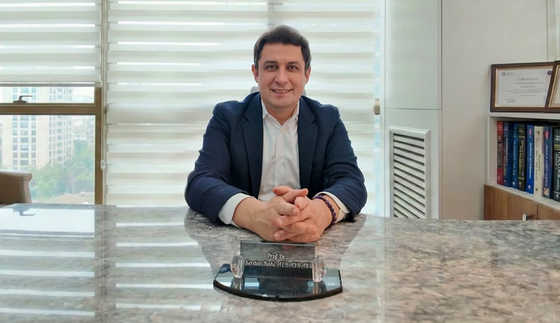
Prof. Dr. Serdar Baki Albayrak
Neurosurgeon · Full Professor of Neurosurgery
Chordoma care starts with a plan that links the biopsy, the operation and the radiotherapy. Prof. Albayrak brings his experience in skull base and spine tumor surgery to each plan, with nerve monitoring during the operation.
After medical school at Marmara University, he trained in neurosurgery at Istanbul University. Clinical fellowships at the University of Helsinki and Harvard Medical School followed.
27+Years
5,000+Operations
64Countries
33Publications
Figures from Prof. Albayrak's professional profile, September 2026. See his full publication list.
Request an AppointmentFrequently Asked Questions
Is chordoma a brain tumor?
Not exactly, since a skull base chordoma grows in the bone under the brain, not in the brain itself. Still, it presses on the brainstem and nerves, so neurosurgeons treat it. For tumors inside the brain, see our brain tumors guide.
Can children get chordoma?
Rarely. MedlinePlus notes that about 5 percent of cases occur in children, mostly at the skull base. For other tumors in children, see our guide to pediatric brain tumors.
Which doctor treats chordoma?
Neurosurgeons operate on skull base and spine chordomas. In the sacrum, orthopedic, general and plastic surgeons often join the team. A radiation oncologist then plans the radiotherapy.
How much does chordoma surgery cost in Turkey?
The cost depends on the location, the route, the hospital stay and any fixation hardware. For example, an endoscopic skull base operation usually costs less than a sacrectomy with fixation. So ask for the cost of your own plan at the consultation, and see our guide to costs.
Prof. Albayrak's Published Research
Prof. Albayrak has no paper on chordoma itself. These co-authored papers cover the surgery of other malignant bone and spine tumors and the control of tumor removal during surgery. Each link opens PubMed; his profile lists all 33 publications.
- Concomitant existence of giant epidural supra and infratentorial metastatic osteosarcoma
- A case of intra-dural malignant peripheral nerve sheath tumor in thoracic spine associated with neurofibromatosis type 1
- Intra-operative magnetic resonance imaging in neurosurgery
Public health information
- National Cancer Institute. Chordoma.
- MedlinePlus Genetics, US National Library of Medicine. Chordoma.
- National Cancer Institute. External Beam Radiation Therapy for Cancer.
- NHS. Bone cancer.
- NHS. Treatment for bone cancer.
- NHS. Double vision.
- NHS. DVT (deep vein thrombosis).
- UK Civil Aviation Authority. Surgical conditions: guidance for health professionals.
- US Centers for Disease Control and Prevention. Medical tourism.
This guide gives general medical information and does not replace a consultation. So diagnosis and treatment decisions need an examination by your own doctor.
Last updated: 15 October 2026. Website editor: Prof. Dr. Serdar Baki Albayrak's clinic, [email protected].
Plan Your Chordoma Consultation in Istanbul
Request an appointment with Prof. Dr. Serdar Baki Albayrak. The team then helps you plan the dates of your visit.
Request an Appointment