
Paid online consultations are available by appointment. For booking, please contact us via Whatsapp.

What Is Ependymoma Surgery?
Ependymoma surgery removes a tumor that grows from the cells lining the brain's fluid spaces or the center of the spinal cord. For most people, it is the first treatment, and complete removal is the single strongest factor for long-term control.
First, in adults, most ependymomas sit in the spine. In children, by contrast, most grow inside the head, often near the fourth ventricle.
Many of these tumors have a clear border. So a careful operation can often take out the whole tumor while it protects the nerves around it.
At a Glance
- Who it helpsAdults and children with an ependymoma of the spinal cord, the lower spine or the brain
- Main goalRemove the whole tumor safely and confirm its exact type
- Common typesSpinal ependymoma, myxopapillary ependymoma, subependymoma, brain ependymoma
- After surgeryMany people need no further treatment; some have focused radiotherapy
- Hospital stayOften about five to ten days, longer after a large operation
- Follow-upRegular brain and spine MRI for many years
Where Are You in Your Decision?
Pick the line that sounds most like you. Each one then opens the matching part of this guide.
- 01My MRI report says ependymoma. Which type is it?Four main types and the usual plan for each
- 02Do I really need surgery, or can radiation do?All treatment options side by side
- 03My tumor is in the spinal cord or the lower spineHow spinal and myxopapillary surgery works
- 04My tumor is in the brainFourth ventricle and upper brain ependymoma
- 05I want to know about risks, recovery and recurrenceWarning signs, timeline and MRI schedule
- 06I am planning surgery from abroadLength of stay, flights and follow-up at home
Types of Ependymoma and What They Mean for Surgery
The type and location of the tumor shape the whole plan. In fact, the NCI notes that location and molecular features predict the outlook better than grade alone.
| Type | Where it grows | Who gets it | Usual plan |
|---|---|---|---|
| Spinal ependymoma (grade 2) | Inside the spinal cord, often in the neck or upper back | Mostly adults | Complete removal; often no further treatment |
| Myxopapillary ependymoma (grade 2) | At the lower tip of the cord, among the nerve roots | Mostly adults | Removal in one piece; radiotherapy if part stays |
| Subependymoma (grade 1) | The wall of a brain ventricle, sometimes the spine | Mostly middle-aged and older adults | MRI checks if small; surgery if it blocks fluid or grows |
| Brain ependymoma (grade 2 or 3) | Fourth ventricle or the upper brain | Children more often; also adults | Maximal safe removal, then usually focused radiotherapy |
Spinal (intramedullary) ependymoma
This tumor grows in the center of the cord and also pushes nerve fibers aside. So it often causes slow back or neck pain, then numbness or weakness. A fluid cavity, or syrinx, may form above or below it; see syringomyelia.
Myxopapillary ependymoma
This type, however, starts on the filum terminale, a thin thread at the lower end of the cord. For example, low back pain, sciatica-like leg pain and, later, bladder problems are typical. Removal in one piece, without opening its capsule, lowers the risk of spread.
Brain ependymoma
Near the fourth ventricle, the tumor can block the flow of brain fluid. Then morning headache, vomiting, dizziness and unsteady walking follow. In the upper brain, by contrast, seizures or weakness on one side may come first.
Several tumors and NF2
Most ependymomas occur by chance and do not run in families. However, people with neurofibromatosis type 2 (NF2) can develop several spinal ependymomas. For them, the plan usually treats the tumor that causes symptoms and watches the rest.
Questions at this stage
Is ependymoma benign or malignant?
Grade 1 and grade 2 ependymomas are low grade, so they grow slowly. Grade 3 tumors, however, are malignant and grow faster. Even a low-grade tumor can harm the cord or brain, so it still needs expert treatment.
How is a spinal ependymoma different from an astrocytoma?
Both grow inside the cord. An ependymoma, however, usually has a clear border, while an astrocytoma tends to blend into the cord. So complete removal is far more often possible with ependymoma.
When to See a Doctor About Ependymoma Symptoms
Slow symptoms deserve a prompt appointment and an MRI. Sudden loss of strength or of bladder control, however, needs an emergency department the same day.
Book a specialist visit soon if you notice:
- Back or neck pain that worsens at night or lasts for weeks
- Numbness, tingling or burning in the arms, legs or trunk
- Leg pain like sciatica without a clear disc problem
- Headaches that wake you or come with vomiting
- Growing clumsiness or unsteady walking
Go to the nearest emergency department (911, 999 or 112) for:
- Sudden weakness in the legs or arms
- New loss of bladder or bowel control
- Numbness in the groin or inner thighs
- Severe headache with repeated vomiting or drowsiness
- Double vision, confusion or a first seizure
Why Speed Matters
In fact, nerve function before surgery strongly predicts function after it. So a weakness that has just begun has a better chance to recover than one that has lasted for months.
Ependymoma Treatment: Surgery, Radiation and Other Options
The NCI lists surgery as the first treatment for an ependymoma whenever it is possible. Radiation, radiosurgery and medicine then play a supporting role.
| Option | Who it suits | Main goal | Limits |
|---|---|---|---|
| Microsurgical removal | Almost everyone whose health allows surgery | Remove the whole tumor and confirm its type | Some tumors stick to vital tissue |
| Focused radiotherapy | Grade 3 tumors; grade 2 tumors with tumor left behind | Control cells that surgery could not reach | Does not replace removal; weeks of daily sessions |
| Stereotactic radiosurgery | Small leftover or returning tumors | Treat a small spot in one to a few sessions | Unsuitable for large tumors or those next to the cord |
| Chemotherapy or trials | Recurrence after surgery and radiation; spread | Slow further growth | Effect on ependymoma remains modest |
| MRI checks only | A small subependymoma without symptoms | Avoid surgery while the tumor stays still | Surgery follows if it grows or blocks fluid |
Why Complete Removal Matters Most
First, a tumor that comes out whole is far less likely to return. For this reason, Prof. Albayrak aims for maximal safe removal, which means taking all the tumor that he can take without new damage.
Also, surgery gives tissue for the final diagnosis. Today, the pathology report should name the molecular subtype as well as the grade, because that subtype guides any further treatment.
Can ependymoma be cured?
Often, yes. Many spinal and myxopapillary ependymomas never return after complete removal. A grade 3 tumor or one with tumor left behind, by contrast, calls for long-term control with radiotherapy and MRI checks.
Do I need radiation after ependymoma surgery?
Not always. After complete removal of a spinal grade 2 tumor or a subependymoma, many people need only MRI checks. Grade 3 tumors and tumors with a remnant, however, usually receive focused radiotherapy.
How Spinal Ependymoma Surgery Works
Spinal ependymoma surgery takes place under general anesthesia, with the patient face down. It often lasts several hours, since the surgeon works slowly between nerve fibers.
- Opening the spine: first, the surgeon lifts the back muscles and removes or hinges the bony arches over the tumor.
- Watching the nerves: then nerve monitoring checks the pathways for movement and feeling throughout the operation.
- Reaching the tumor: after the dura opens, a short cut along the midline of the cord exposes the tumor.
- Removing it: under the microscope, the surgeon finds the border and then separates the tumor from the cord.
- Closing: the dura closes watertight, and the bony arches also go back where the plan allows.
Prof. Albayrak uses microsurgery and neuronavigation for these tumors. His 2004 first-author review also describes how imaging during surgery can guide tumor removal.
Myxopapillary Ependymoma Surgery
Here the tumor hangs among the nerve roots of the cauda equina. So the surgeon frees it from the roots, then cuts the filum thread above and below it. Then the tumor comes out in one piece.
If the capsule breaks, however, tumor cells can spread through the spinal fluid. For this reason, a large tumor that has broken open often receives radiotherapy afterward.
Tests Before and After Surgery
Before surgery, an MRI of the whole brain and spine looks for spread along the fluid pathways. After surgery, Prof. Albayrak often checks a spinal fluid sample, taken at least two weeks later, for tumor cells.
For other tumors of the spine, see our guide to spinal cord and spine tumors.
Will I be able to walk after spinal ependymoma surgery?
Most people who walk before surgery also walk afterward. Many, however, feel more numbness or weakness for a few weeks, and then improve with rehabilitation. Your nerve function before surgery is the best guide to the result.
Do I need screws or a fusion after spinal ependymoma surgery?
Usually not in adults. The bony arches often go back in place, which helps keep the spine stable. Long-term X-rays, however, check the spine's shape, especially after surgery over many levels.
Brain Ependymoma Surgery
For a tumor near the fourth ventricle, the surgeon goes in through the back of the head, below the cerebellum. In the upper brain, a craniotomy above the tumor gives the shortest safe route.
Tumors of the fourth ventricle often reach toward the brainstem and the nerves for swallowing and eye movement. So Prof. Albayrak uses nerve monitoring to protect these nerves. He also uses microsurgery and fluorescence-guided techniques to see the tumor border.
When the Fluid Pathway Gets Blocked
Also, a blocked fourth ventricle can raise pressure in the head. In that case, he first restores fluid flow with an endoscopic third ventriculostomy or a shunt; our hydrocephalus treatment guide explains both.
For the operation itself, see our guide to cerebellar tumor surgery. Our brain tumors page covers other tumor types.
How long does ependymoma surgery take?
Often four to eight hours, depending on the size and site of the tumor. Nerve monitoring and slow microsurgery take time. So the length of the operation says little about its difficulty.
Ependymoma Surgery Risks and Success Rate
The main risks come from the delicate tissue around the tumor. Most effects are temporary, but some can last.
- New numbness or weakness: common for a few weeks after spinal surgery; most of it improves with rehabilitation.
- Bladder, bowel or sexual changes: also possible after lower spine surgery; they often ease over months.
- Spinal fluid leak: a clear wound discharge or headache when you stand up; see our guide to CSF leak repair.
- Swallowing or eye movement problems: after fourth ventricle surgery, usually temporary.
- Infection, bleeding and blood clots: uncommon, as with other major operations.
- Spinal deformity: mainly in children and after surgery over many levels.
What the Numbers Say
The NCI reports a five-year relative survival of about 88 percent for ependymoma overall. However, grade, type, location, age and the amount of tumor left all change that figure. So ask your surgeon to explain your own situation.
What is the success rate of ependymoma surgery?
For spinal and myxopapillary tumors with a clear border, complete removal is often possible. Brain tumors near the brainstem, however, may leave a small part behind. Your surgeon can judge your chances from your MRI and examination.
What are the long-term effects of ependymoma surgery?
Many people return to work and normal life. Some, however, keep mild numbness, stiffness or bladder changes. Radiotherapy can add tiredness and, in the brain, effects on memory over time.
Ependymoma Surgery Recovery Time
Most people stand up within a day or two and leave the hospital within about a week to ten days. After that, recovery of nerve function often continues for several months.
| Stage | What happens | Your part |
|---|---|---|
| First 48 hours | Close watch, pain relief, early MRI | Lie flat if advised; move your feet and hands |
| Days 3 to 10 | First walks with a physiotherapist; discharge | Walk a little more each day |
| Weeks 2 to 3 | Stitches out; pathology report with molecular subtype | Discuss the result and any radiotherapy |
| Weeks 2 to 6 | Rehabilitation; spinal fluid test if planned | No heavy lifting or twisting; keep up exercises |
| Months 2 to 6 | Nerve function keeps improving; first follow-up MRI | Return to desk work when your surgeon agrees |
Follow-Up MRI and Recurrence
Ependymoma can return, most often within the first two to five years and at the original site. So Prof. Albayrak's usual plan is an MRI of the brain and spine every three to four months for two years. After that, scans come every six months until year five, then once a year.
Also, a recurrence found early is often small. Then a second operation, radiosurgery or radiotherapy can usually treat it.
Ependymoma in Children
Children, however, more often have brain ependymomas, and their plan involves a pediatric oncology team. Read our guides to brain tumors in children and spinal cord tumors in children.
How long does it take to recover from spinal ependymoma surgery?
Many people feel well enough for daily life within six weeks. Walking, balance and bladder control, however, can keep improving for six months or more. Rehabilitation speeds this up.
What are the signs of ependymoma recurrence?
Often there are none at first, which is why regular MRI matters. Later, the old symptoms may return, such as pain, numbness, weakness or headache. Report any such change before your next planned scan.
Coming to Istanbul from the US, Canada, the UK or Europe
Ependymoma surgery rarely counts as an emergency, so it suits a trip you arrange ahead. Sudden weakness or loss of bladder control, however, calls for care at home first.
How Long to Stay in Istanbul
First, count the consultation, any new MRI and about a week to ten days in hospital. Then add a few days until your surgeon clears you to fly. So many people stay about two to three weeks in total.
When You Can Fly Home
The UK CAA advises waiting about seven days after surgery inside the skull. For spine surgery, it sets no fixed period, so your surgeon decides at the check-up.
Also, the US CDC warns that flying soon after surgery raises the risk of blood clots. So walk in the aisle and drink water on the flight.
Follow-Up at Home
Before you leave, ask for the operation report, the discharge summary and copies of your scans. The final pathology report, with its molecular subtype, follows by email. If you need radiotherapy, your local oncology team can then plan it near home.
For insurance and Medicare, see planning surgery in Turkey from the US. For NHS rules and travel insurance, see planning from the UK. From Canada, check with your provincial health plan and private insurer before you book.
Your treatment in Istanbul
From Your First Message to Flying Home
Treatment in Istanbul follows four clear steps. Each one answers a practical question about your trip.
STEP 01
Request an appointment
Use the form on this page, WhatsApp or call +90 532 308 97 72. Then share your main symptoms, where the tumor sits and your travel dates. You can write in English or Turkish.
STEP 02
Consultation and tests in Istanbul
First, Prof. Albayrak examines your strength, feeling and reflexes at his clinic in Teşvikiye. Next, he goes through your MRI scans with you. A fresh MRI of the brain and whole spine often follows in Istanbul.
He then explains whether surgery suits you and what it aims to achieve. Questions worth asking include:
- Can the whole tumor come out safely?
- Which functions could change after surgery?
- Will I need radiotherapy afterward?
What should I bring to the consultation?
First, bring all MRI images on a disc or USB drive, with the written reports. Also bring any earlier operation or pathology reports and a list of your medicines.
STEP 03
Surgery and hospital stay
If you choose surgery, the operation takes place in a hospital in Istanbul. Prof. Albayrak names the hospital at the consultation, along with the likely stay. In addition, a relative or companion can usually stay with you.
STEP 04
Recovery, fitness to fly and follow-up
After discharge, you rest at a nearby hotel and keep walking each day. Then, before the flight, your surgeon checks the wound and confirms that you can fly.
Back home, then, seek urgent help for new weakness, loss of bladder control, fever or a swollen leg.
Can someone travel with me?
Yes, and it helps. A companion can carry bags, since lifting is off limits for weeks. Also, they can watch for warning signs during the first days.
Your Ependymoma Surgeon in Istanbul
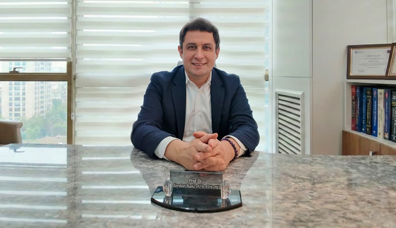
Prof. Dr. Serdar Baki Albayrak
Neurosurgeon · Full Professor of Neurosurgery
Prof. Albayrak removes ependymomas of the spinal cord, the lower spine and the brain with microsurgery and nerve monitoring. He plans each operation around the tumor type and your nerve function.
After medical school at Marmara University, he trained in neurosurgery at Istanbul University. Clinical fellowships at the University of Helsinki and Harvard Medical School followed.
27+Years
5,000+Operations
64Countries
33Publications
Figures from Prof. Albayrak's professional profile, September 2026. See his full publication list.
Request an AppointmentFrequently Asked Questions
Is ependymoma cancer?
Ependymoma is a primary tumor of the brain or spinal cord. Grade 3 tumors count as cancer, while most spinal and myxopapillary tumors are low grade. Ependymomas rarely spread outside the nervous system, the NCI notes.
Is ependymoma hereditary?
Usually not. Most cases arise by chance. NF2, however, raises the risk of spinal ependymomas, so family members may need checks.
Can a recurrent ependymoma have surgery again?
Often, yes. A second operation is a common first step when the tumor returns at the same site. Radiosurgery or radiotherapy may follow, depending on earlier treatment.
How much does ependymoma surgery cost in Turkey?
The cost depends mostly on the tumor site, the length of the operation and the hospital stay. Rehabilitation and any new MRI also add to it. So ask for the cost of your plan at the consultation, and see our guide to costs.
Prof. Albayrak's Published Research
Prof. Albayrak has no paper on ependymoma itself. Instead, the first paper describes a tumor that spread inside the cervical spinal cord and along its lining in an adult. The second covers the removal and return of a tumor inside the dura of the thoracic spine, and the third reviews imaging during tumor surgery.
Each link opens PubMed; his profile lists all 33 publications.
- Cervical leptomeningeal and intramedullary metastasis of a cerebral PNET in an adult
- A case of intra-dural malignant peripheral nerve sheath tumor in thoracic spine associated with neurofibromatosis type 1
- Intra-operative magnetic resonance imaging in neurosurgery
Public health information
- National Cancer Institute, NCI-CONNECT. Ependymoma: Diagnosis and Treatment.
- National Cancer Institute. Adult Central Nervous System Tumors Treatment (PDQ).
- National Cancer Institute. Childhood Ependymoma.
- MedlinePlus, US National Library of Medicine. Brain tumor: primary, adults.
- NHS. Brain tumours.
- NHS. DVT (deep vein thrombosis).
- UK Civil Aviation Authority. Surgical conditions: guidance for health professionals.
- US Centers for Disease Control and Prevention. Medical tourism.
This guide gives general medical information and does not replace a consultation. So diagnosis and treatment decisions need an examination by your own doctor.
Last updated: 24 October 2026. Website editor: Prof. Dr. Serdar Baki Albayrak's clinic, [email protected].
Plan Your Ependymoma Consultation in Istanbul
Request an appointment with Prof. Dr. Serdar Baki Albayrak. The team then helps you plan the consultation, any tests and your stay.
Request an Appointment