
Paid online consultations are available by appointment. For booking, please contact us via Whatsapp.

What Is a Parietal Lobe Tumor?
A parietal lobe tumor grows in the upper side of the brain, just behind the frontal lobe and under the crown of the head. This area handles touch, body position, direction and left-right sense. So the first sign is often numbness or clumsiness rather than a headache.
Each side of the brain has its own parietal lobe, but the two sides do different jobs. Our overview of brain tumor treatment also covers tumors in other areas.
Surgery forms the main treatment for most of them. The aim is to remove as much tumor as is safe while protecting touch, language and vision. In addition, removal often eases tumor-related seizures.
At a Glance
- Where it sitsThe upper side of the brain, behind the frontal lobe and above the ear
- First signNumbness, a tingling seizure or a clumsy hand on the opposite side
- Common typesGlioma, meningioma and metastasis from cancer elsewhere
- Main treatmentMicrosurgery that removes as much tumor as is safe
- Key decisionLeft or right side, and how close the tumor lies to touch, language and vision pathways
- Hospital stayUsually 3 to 5 days, then a few weeks of recovery
Which Symptom Brought You Here?
First, pick the line closest to your situation. It then takes you to the matching part of this guide.
- 01Numbness, tingling spells or a clumsy handSigns from the touch area
- 02New trouble writing, reading or doing sumsSigns of a left-side tumor
- 03A relative ignores the left side or gets lostSigns of a right-side tumor
- 04The MRI shows a mass in the parietal lobeWhich tumor it might be
- 05Unsure which operation fitsTreatment options compared
- 06Planning surgery from abroadLength of stay, flights and care back home
Parietal Lobe Tumor Symptoms
The most common symptoms of a parietal lobe tumor are numbness on the opposite side, trouble recognizing objects by touch and short tingling seizures. Then, depending on the side, language or attention problems may follow. As the tumor grows, headaches can join in.
Numbness, tingling and clumsy hands
The front strip of the parietal lobe receives touch, pain, heat and position signals from the body. So a tumor here dulls feeling in the hand, arm or leg on the other side.
For example, with closed eyes you may fail to tell a key from a coin in your pocket. Buttons and small objects also get harder, even though muscle strength stays normal. Reaching for a cup without looking can miss the target, too.
Tingling seizures
Parietal seizures usually start with a feeling rather than jerking. Tingling, burning or an electric sensation begins in one finger, then spreads up the arm or to the face within seconds to minutes.
Sometimes the spell reaches the movement strip next door, and the arm starts to jerk. So repeated tingling spells that always start in the same place call for a brain MRI.
Left side: writing, sums and words
In most people, the lower left parietal lobe supports reading, writing, numbers and finger naming. A tumor there can make texting, paying a bill or simple sums suddenly hard.
Doctors call one rare cluster Gerstmann syndrome: trouble writing, trouble with sums, confusing fingers and mixing up left and right. All four seldom appear together, but even one new sign deserves a check.
Right side: neglect and getting lost
The right parietal lobe spreads attention across both sides of space and the body. So a tumor here can make a person ignore their left side. For example, they eat only the right half of the plate or bump the left door frame.
Often, the person does not notice the problem at all, a state doctors call anosognosia. Family members therefore tend to see the change first. Getting lost on familiar routes and trouble dressing are common clues, too.
Vision gaps and headaches
Vision fibers run deep under the parietal lobe. A tumor can therefore dim the lower part of vision on the opposite side, so stairs and kerbs seem to vanish.
Later, swelling around the tumor raises pressure in the skull. Headaches then grow worse, especially in the morning. Our guide to brain tumour symptoms covers pressure signs in detail.
Questions at this stage
Is numbness in the hand always a sign of a brain tumor?
No, because carpal tunnel syndrome and neck nerve problems cause it far more often. However, numbness of the face, arm and leg on the same side, or numbness that comes in spells, calls for a brain MRI.
What are the first signs of a right parietal lobe tumor?
Often the family notices them first. The person ignores things on the left, gets lost on known routes or struggles to dress. Numbness or tingling on the left side of the body may also appear.
What are the symptoms of a left parietal lobe tumor?
Typical signs include numbness on the right side, new trouble with writing or sums and mixing up left and right. Reading and word finding can also slow down. Unlike right-side tumors, most people notice these changes themselves.
When to See a Doctor
Most parietal lobe tumor symptoms build over weeks, so you usually have time to book a visit. Some signs, however, call for emergency care.
Book a doctor's visit if you notice:
- Numbness or clumsiness on one side that grows over weeks
- Repeated tingling spells that start in the same place
- New trouble with writing, reading or simple sums
- A relative who ignores the left side or gets lost on known routes
Seek emergency care for:
- Sudden weakness or numbness of the face, arm or leg
- Sudden trouble speaking or understanding speech
- A first seizure, or a seizure that lasts over 5 minutes
- A severe headache with vomiting, or growing drowsiness
Sudden signs can mean a stroke or rising pressure in the skull. So go to the nearest emergency department first, rather than waiting for a visit abroad.
Why the Exact Spot Matters
The parietal lobe has several parts with different jobs. So the exact spot of the tumor shapes both the symptoms and the surgical plan.
| Part of the parietal lobe | Main job | Typical sign from a tumor |
|---|---|---|
| Front strip (touch cortex) | Touch, pain, heat and position sense | Numbness, tingling, sensory seizures on the other side |
| Upper part | Guiding the hand to a target, body map | Missing the target when reaching without looking |
| Lower left part | Writing, numbers, finger naming, left-right sense | Trouble writing, reading or doing sums |
| Lower right part | Attention, spatial sense, awareness of the left side | Ignoring the left side, getting lost, trouble dressing |
| Deep white matter | Carrying vision fibers to the back of the brain | Loss of lower vision on the other side |
| Inner surface (precuneus) | Sense of self, recalling scenes | Often silent until the tumor grows large |
Left Versus Right Parietal Lobe Tumor
The two sides do not share the same work. So the side of the tumor changes the signs, the mapping before surgery and the focus of rehabilitation.
| Feature | Left parietal tumor | Right parietal tumor |
|---|---|---|
| Numb side | Right face, arm and leg | Left face, arm and leg |
| Language | Trouble reading, writing and finding words | Usually spared |
| Numbers and fingers | Trouble with sums and naming fingers | Usually spared |
| Attention | Marked neglect stays rare | Ignoring the left side is common |
| Awareness of the problem | The person usually notices | The person often denies it |
| Focus of surgery | Language mapping, sometimes awake | Protecting attention, touch and vision fibers |
Which Tumors Grow in the Parietal Lobe?
Gliomas, meningiomas and metastases form the main groups. Also, because the parietal lobe has one of the widest surfaces in the brain, metastases from cancer elsewhere settle here often.
| Tumor type | Starts from | Behavior | Goal of surgery |
|---|---|---|---|
| Glioblastoma | Brain support cells | Malignant, fast growing | Maximal safe removal, then further treatment |
| Astrocytoma and oligodendroglioma | Brain support cells | Often slower; seizures common in young adults | The widest safe removal |
| Meningioma | The brain lining | Mostly benign; pushes the brain from outside | Full removal with its attachment |
| Metastasis | Lung, breast, kidney or skin cancer | Depends on the source; marked swelling | Removal of a single, reachable spot |
| Lymphoma | Immune cells | Malignant | Usually a biopsy for diagnosis only |
Only tissue tests confirm the exact type. For details by type, see our guides to astrocytoma, oligodendroglioma, meningioma and brain metastases.
Is a parietal lobe tumor cancerous?
Some are and some are not. Meningiomas in this area are mostly benign, and full removal often cures them. Gliomas and metastases, however, also occur often, so the pathology report gives the final answer.
What is the most common parietal lobe tumor?
In adults, gliomas lead, together with meningiomas and metastases. For people with a known cancer, however, a new parietal mass often turns out to be a metastasis. In children, gliomas account for most parietal tumors.
Tests Before Parietal Lobe Tumor Surgery
An MRI with contrast confirms a mass in the parietal lobe. Further tests then map the brain around it, so the surgeon can plan a safe route.
- MRI with contrast: shows the position, the size and any swelling
- Perfusion and spectroscopy MRI: give clues about how active the tumor looks
- Functional MRI: locates the touch, movement and language areas
- Tractography (DTI): maps the vision and movement fibers around the tumor
- Visual field test: finds a lower vision gap and gives a baseline
- Neuropsychological tests: measure attention, language, sums and spatial sense
- EEG: records seizure activity and helps with medicine choices
However, only pathology confirms the tumor type. So the sample comes either from the removed tumor or, when needed, from a needle biopsy.
Can a parietal lobe tumor be mistaken for a stroke?
Yes, because both cause numbness on one side. A stroke, however, starts within minutes, while a tumor builds over weeks. An MRI tells them apart quickly, so sudden signs always need emergency care first.
Parietal Lobe Tumor Treatment Options Compared
Microsurgery that removes as much tumor as is safe forms the main treatment. Radiotherapy and drug treatment then follow, depending on the pathology result. For benign tumors removed fully, MRI checks alone often suffice afterward.
Prof. Albayrak first maps how close the tumor lies to the touch strip, the language network and the vision fibers. That map then decides the type and the extent of surgery.
| Option | Who it may suit | Aim |
|---|---|---|
| Full removal | Meningiomas and tumors with clear borders | Remove the tumor without a trace, prevent regrowth |
| Widest safe removal | Gliomas and tumors near key areas | Relieve pressure and prepare for further treatment |
| Awake surgery | Lower left tumors near the language and number network | Protect speech, reading and sums |
| Needle biopsy | Deep or multiple tumors, or suspected lymphoma | Confirm the diagnosis |
| Radiosurgery | Small metastases, or people unfit for surgery | Focused radiation, run by radiation oncology |
Can a parietal lobe tumor be removed?
Most of them can, because the parietal lobe lies close to the skull surface. The real question concerns how much can come out safely. That depends on the tumor type and its distance from the touch, language and vision pathways.
Parietal Lobe Tumor Surgery: What the Surgeon Does
The surgeon opens a window in the skull right over the tumor. Under the operating microscope, he then separates the tumor from the brain around it and removes it.
Throughout, neuronavigation shows the position of each instrument in the brain. Nerve monitoring also tracks the touch and movement pathways without a break.
The operation usually takes 3 to 6 hours, and longer with awake mapping. Our brain tumor surgery overview explains the general steps.
Awake Surgery Near Language Areas
When the tumor lies in the lower left parietal lobe, close to the language and number network, Prof. Albayrak may advise awake surgery. During the key stage, you name objects, read or count.
So he can stop at the point where a function starts to falter. Our awake craniotomy guide explains each stage.
Protecting Touch and Vision
The touch strip sits right next to the movement strip. So nerve monitoring warns the team early if either pathway comes under strain. Tractography also shows the route of the vision fibers, and the surgeon plans the approach around them.
How long does parietal lobe tumor surgery take?
It usually takes 3 to 6 hours, depending on the size and position of the tumor. Awake surgery and detailed mapping take longer. Time in the recovery unit then adds a few more hours.
Can a parietal lobe tumor be removed completely?
Often, yes, especially meningiomas with clear borders. Gliomas, however, spread into nearby brain, so some cells usually stay behind. For that reason, follow-up scans continue after any glioma surgery.
Risks of Parietal Lobe Tumor Surgery
The main risks specific to this surgery involve touch, lower vision, language and seizures. Most of these effects stay mild or improve over weeks. Like any brain operation, it also carries a risk of bleeding and infection.
| Risk | Why it can happen | How the team lowers it |
|---|---|---|
| Short-term numbness or clumsiness | Swelling around the touch strip | Nerve monitoring, swelling treatment, early physiotherapy |
| Lower vision gap | Strain on the deep vision fibers | Tractography shows the fiber route in the plan |
| Trouble with words, writing or sums | Strain on the lower left network | Awake surgery and function mapping |
| Seizures | Irritation of the brain surface | Seizure medicine before and after surgery |
| Bleeding and infection | General risks of any brain surgery | Careful vessel control, sterile technique, antibiotics |
Your own risk depends on the tumor type, its exact spot and your general health. So the surgeon discusses possible losses openly with you and your family before surgery.
Will the numbness go away after surgery?
Numbness that increases after surgery usually fades within weeks as swelling settles. Numbness present before surgery, in contrast, improves partly or fully, depending on the tumor type and how long it lasted. Full recovery can take 6 to 12 months.
Recovery After Parietal Lobe Tumor Surgery
Most people stay in hospital for 3 to 5 days and return to daily tasks within a few weeks. Touch and spatial sense, however, can take months to recover.
A Typical Timeline
| When | What usually happens |
|---|---|
| First 24 hours | Close watch in intensive care, with checks of alertness and limb movement |
| Days 2-5 | Standing, walking, a control MRI and discharge |
| Week 2 | Stitch removal and a talk about the pathology result |
| Weeks 3-6 | Return to desk work; radiotherapy starts, if needed |
| Months 3-6 | Clear gains in touch and fine hand skills |
| Months 6-12 | The lasting picture becomes clear; regular MRI checks |
Rehabilitation, Neglect and Driving
Occupational therapy matters most for neglect and spatial problems. For example, scanning tasks teach the eyes to search the left side on purpose. Texture exercises with the fingers also speed up the return of touch.
People with seizures or a vision gap should not drive. UK drivers must tell the DVLA about a brain tumor, and the time off the road depends on the tumor and treatment. Elsewhere, check your local licensing rules.
Does neglect of the left side get better?
In most people, neglect eases clearly over weeks and months. Occupational therapy and exercises that draw attention to the left speed this up. Even so, a mild neglect can remain in some people.
Can a parietal lobe tumor affect walking?
Yes, when position sense in the leg fails, walking without looking at the floor gets hard. A lower vision gap can also make you miss steps. Physiotherapy usually reduces both problems clearly.
Parietal Lobe Tumor or Something Else?
Parietal tumor signs often get mistaken for stroke, a trapped nerve in the neck, carpal tunnel syndrome or dementia. All of them can cause numbness, clumsiness or getting lost. A few clues, however, help tell them apart.
| Condition | Onset | Pattern of numbness | Telling clue |
|---|---|---|---|
| Parietal lobe tumor | Weeks to months, gradual | Face, arm and leg on one side | Touch recognition loss, neglect, seizures, mass on MRI |
| Stroke | Minutes, sudden | Face, arm and leg on one side | Sudden start, vascular risk factors |
| Neck nerve root | Days, with pain | A strip down the arm | Neck pain spreading to the arm, face spared |
| Carpal tunnel | Slow, worse at night | The first three fingers | Night waking, eases with shaking the hand |
| Dementia | Years, both sides | No numbness | Memory loss leads, no one-sided signs |
A strip of arm pain and numbness points more toward the neck, as our cervical disc herniation guide explains. Frontal tumors, in contrast, change personality and movement, while temporal tumors affect memory. See our guides to frontal lobe tumors and temporal lobe tumors.
Coming to Istanbul from the US, Canada, the UK or Europe
Parietal lobe tumor surgery needs one trip for the consultation, tests, surgery and early recovery. Further treatment, if needed, then usually continues at home.
How Long to Stay in Istanbul
First, add up the consultation, any new mapping scans, the operation and a hospital stay of 3 to 5 days. Then rest near the hospital until the stitches come out. Overall, many plans come to about two weeks in Istanbul.
When You Can Fly Home
UK aviation guidance advises waiting about 7 days after brain surgery before flying. Air left inside the skull can expand at altitude. The US CDC also warns that flying soon after surgery raises the risk of blood clots.
Follow-Up at Home
Before you leave, ask for the discharge summary, the pathology report and copies of all scans. Then share them with your local doctor, and book occupational therapy if neglect or clumsiness remains.
For insurance and Medicare, see brain surgery in Turkey: planning from the US. For NHS rules and travel insurance, see planning from the UK. From Canada, check with your provincial health plan and private insurer before you book.
Your treatment in Istanbul
From Your First Message to Flying Home
Your treatment in Istanbul follows four clear steps. Each step then answers one practical question.
STEP 01
Request an appointment
Use the form on this page, WhatsApp or call +90 532 308 97 72. A short message is enough: your diagnosis, your country and your travel dates.
You can write in English or Turkish, so use whichever feels easier. The team then agrees the consultation date with you.
STEP 02
Consultation and tests in Istanbul
First, Prof. Albayrak examines you at his clinic in Teşvikiye. Next, he goes through your MRI scans with you. If needed, mapping scans, a visual field test or neuropsychological tests can follow in Istanbul.
He then explains whether surgery suits you, and which approach fits. Questions worth asking include:
- How much of the tumor can come out safely?
- Will I need awake surgery?
- What does the side of the tumor mean for touch, language and vision?
What should I bring to my parietal lobe tumor consultation?
First, bring your MRI scans on a disc or USB drive, with the written reports. Also bring a list of your medicines and any EEG or vision test results. A relative who has seen the changes helps, too.
STEP 03
Surgery and hospital stay
If you choose surgery, the operation takes place in a hospital in Istanbul. Prof. Albayrak then names the hospital at the consultation, along with the likely stay.
STEP 04
Recovery, fitness to fly and follow-up
After discharge, you rest at a nearby hotel, ideally with a companion. Then, before the flight, your surgeon checks the wound and confirms that you can fly.
Back home, get urgent help for any of these signs:
- A seizure, especially a long or repeated one
- A severe or worsening headache, or repeated vomiting
- New weakness, confusion or trouble speaking
- Fever, redness or fluid leaking from the wound
Can I start rehabilitation at home after surgery in Istanbul?
Yes, in most cases. Your local therapist then works from the discharge summary and the notes about touch, vision and attention. So book that first session before you travel, if you can.
Your Parietal Lobe Tumor Surgeon in Istanbul
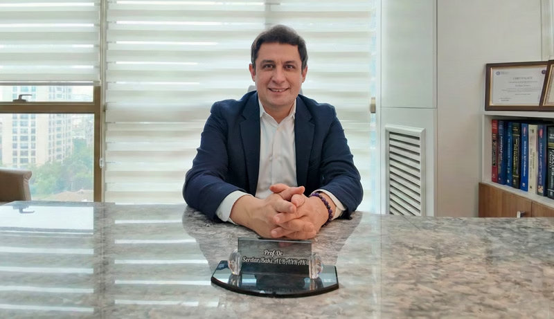
Prof. Dr. Serdar Baki Albayrak
Neurosurgeon · Full Professor of Neurosurgery
A parietal lobe tumor sits close to touch, language and vision pathways, so the plan starts with careful mapping. For this reason, Prof. Albayrak combines microsurgery with navigation, nerve monitoring and, near language areas, awake surgery.
He completed his neurosurgery training at Istanbul University in 2004. He then took clinical fellowships at the University of Helsinki and Harvard Medical School. At Harvard, he trained at Brigham and Women's Hospital.
27+Years
5,000+Operations
64Countries
33Publications
Figures from Prof. Albayrak's professional profile, September 2026. See his full publication list.
Request an AppointmentFrequently Asked Questions
Does a parietal lobe tumor cause headaches?
Yes, but headache is rarely the first sign. As the tumor and its swelling grow, pressure in the skull rises. The pain then tends to peak in the morning and with coughing or bending.
Do children get parietal lobe tumors?
Yes, although brain tumors in children more often grow in the cerebellum and brainstem. Most parietal tumors in children turn out to be gliomas. New hand clumsiness or a seizure in a child always deserves a check, as our pediatric brain tumors guide explains.
Which specialists treat a parietal lobe tumor?
A neurosurgeon leads the diagnosis and surgery. A neurologist manages seizures, and a neuropsychologist tests attention and language. For malignant tumors, oncology and radiation oncology join the team.
How much does parietal lobe tumor surgery cost in Turkey?
The cost depends on the operation, the hospital stay and the tests you need. Mapping scans, awake surgery and your hotel stay also count. So ask for the cost of your own plan at the consultation, before you decide.
Prof. Albayrak's Published Research
None of Prof. Albayrak's papers focuses on parietal lobe tumors alone. The closest cover MRI during tumor surgery, a metastasis that mimicked a glioma in the cerebral hemisphere and the origin of meningiomas. Each link opens the record; his profile lists all 33 publications.
- Intra-operative magnetic resonance imaging in neurosurgery
- Cerebral metastasis of small-cell lung carcinoma mimicking a supratentorial cystic astrocytoma
- The Origin of Meningiomas
Public health information
- National Cancer Institute. Adult Central Nervous System Tumors Treatment (PDQ), patient version.
- MedlinePlus, US National Library of Medicine. Brain tumor - primary - adults.
- MedlinePlus, US National Library of Medicine. Metastatic brain tumor.
- NHS. Brain tumours.
- National Institute for Health and Care Excellence. Brain tumours (primary) and brain metastases in over 16s (NG99): recommendations.
- UK Civil Aviation Authority. Surgical conditions: guidance for health professionals.
- US Centers for Disease Control and Prevention. Medical tourism.
- GOV.UK. Brain tumour and driving.
This guide gives general medical information and does not replace a consultation. So diagnosis and treatment decisions need an examination by your own doctor.
Last updated: 2 November 2026. Website editor: Prof. Dr. Serdar Baki Albayrak's clinic, [email protected].
Plan Your Parietal Lobe Tumor Consultation in Istanbul
Request an appointment with Prof. Dr. Serdar Baki Albayrak. The team then helps you plan the dates of your visit.
Request an Appointment