
Paid online consultations are available by appointment. For booking, please contact us via Whatsapp.

What Is a Spinal AVM?
A spinal arteriovenous malformation (spinal AVM) is an abnormal short circuit between arteries and veins in or around the spinal cord. High-pressure blood skips the tiny capillaries and floods the veins, so the cord can no longer drain well.
In practice, doctors use "spinal AVM" as an umbrella term. It covers a tangle of vessels inside the cord and also a single small leak in the cord's covering, the spinal dural arteriovenous fistula (spinal dAVF).
Both types also harm the cord in two ways. First, blood pools and the tissue swells for lack of oxygen. Second, a thin vessel wall can tear and bleed.
At a Glance
- Most common typeSpinal dural AV fistula, mostly in men over 50
- Typical startLeg weakness and bladder trouble that creep up over months
- Sudden formSevere back pain and weakness from a bleed, often in younger people
- Key testSpinal catheter angiography after an MRI
- Main treatmentsMicrosurgery, embolization or both together
- GoalClose the short circuit for good and stop the decline
Where Are You in Your Decision?
Pick the line that sounds most like you. Each one then opens the matching part of this guide.
- 01My legs and bladder keep getting worseSlow and sudden symptoms, and what they mean
- 02Doctors treated me for something elseWhy spinal AVMs pass for stenosis, disc or myelitis
- 03I was told I have a spinal fistula or AVMThe types and how each one behaves
- 04I must choose surgery or embolizationWhat decides the route for your case
- 05I want to know if I will walk betterRecovery time and what usually improves
- 06I am planning treatment abroadRecords to bring, length of stay and flights
Types of Spinal Vascular Malformations
Doctors sort spinal AVMs into four main types by location and blood flow. The type matters too, because it predicts how fast symptoms start, the bleeding risk and the best treatment.
| Type | Where it forms | Who it affects | How it starts | Main treatment |
|---|---|---|---|---|
| Type I: spinal dural AV fistula | In the dura where a nerve root exits | Mostly men over 50 | Slow leg weakness over months | Surgical disconnection or embolization |
| Type II: glomus (intramedullary) AVM | Inside the cord, as a compact tangle | Children and young adults | Often a sudden bleed | Microsurgery, embolization or both |
| Type III: juvenile (diffuse) AVM | Cord, coverings and nearby tissue | Children and teens, very rare | Bleed or growing weakness | Staged embolization, surgery in select cases |
| Type IV: perimedullary AV fistula | On the cord surface, under the covering | Young adults | Growing weakness or a bleed | Surgery or embolization by fistula size |
| Spinal cavernoma | Inside the cord, low-flow cluster | Any age | Small repeated bleeds, step by step | Microsurgery if it causes symptoms |
A cavernoma differs from an AVM, since its blood flows slowly. So our cavernoma guide covers it separately.
Why the dural fistula is the most common type
Overall, the spinal dural AV fistula leads the list of spinal vascular malformations. Experts think it develops during adult life rather than at birth.
Specifically, it sits where a nerve root leaves the spinal canal. There, a tiny artery of the dura joins a vein on the cord surface directly. The fistula itself is small, yet arterial pressure spreads up the whole venous network of the cord.
As a result, the cord swells, and doctors call this venous congestive myelopathy. Most fistulas lie in the lower chest or upper lower back. Still, the first signs often start at the tip of the cord, so the legs and bladder suffer first.
In fact, the same kind of leak can also form in the coverings of the brain. That form, with its very different symptoms, appears in our dural AV fistula guide.
Spinal AVM Symptoms: Slow or Sudden?
Symptoms of a spinal AVM either creep up or strike at once, and the type decides which. A fistula usually worsens over months, while an AVM inside the cord often announces itself with a bleed.
| Feature | Slow course (mostly fistula) | Sudden course (mostly AVM bleed) |
|---|---|---|
| Onset | Over months, quietly | Within minutes or hours |
| First sign | Weak legs, bladder changes | Sudden, severe back pain |
| Age | Usually over 50 | Usually children and young adults |
| Pattern | Ups and downs, but worse overall | Sudden paralysis, then some recovery |
| How urgent | Prompt specialist visit | Emergency department the same day |
Weak, heavy legs that get worse with walking
Stairs get harder, the feet drag and you trip more often. Typically, the legs tire with walking or standing and recover partly with rest. Then numbness, tingling or burning in the legs often joins in.
Exercise, long walks or straining can also make symptoms flare. This up-and-down pattern is an important clue to a vascular cause.
Bladder, bowel and sexual changes
Urgency, leaking urine or constipation can start early too. In men, erection problems are sometimes the very first sign. So these changes deserve attention even when walking still feels fine.
Sudden pain and weakness from a bleed
A bleed causes a sudden, stabbing pain in the back. Then, within minutes or hours, the legs grow weak or numb. Therefore, this needs emergency care.
Questions about symptoms
Can a spinal AVM cause back pain?
Yes. Back pain is common and can spread into the legs, which is why many people first think of a slipped disc. Sudden, severe back pain, however, can mean a bleed and needs urgent care.
Does pregnancy affect a spinal AVM?
It can. Blood volume and pressure in the abdomen rise in pregnancy, so some malformations cause symptoms for the first time. New leg weakness or sudden back pain in pregnancy therefore needs a doctor at once.
When to See a Doctor
Slow symptoms need a prompt, planned visit, because every month of congestion can cost function. Sudden symptoms, in contrast, need emergency care the same day.
Book a specialist visit if you notice:
- Leg weakness or numbness that grows over weeks or months
- New bladder urgency, leaking or constipation with leg symptoms
- Walking problems that persist after stenosis or disc treatment
- Spinal cord swelling on MRI without a clear cause
Go to the nearest emergency department (911, 999 or 112) for:
- Sudden, stabbing pain in the back or neck
- Weakness or numbness in the legs within hours of that pain
- Sudden loss of bladder control, or you cannot pass urine
- A known spinal AVM with suddenly worse symptoms
A spinal cord bleed needs local emergency care first. So never wait to see if it settles, and do not plan a trip abroad before that care.
Why Spinal AVMs Often Get Diagnosed Late
A spinal AVM mimics much more common spine problems, so the true cause can hide for months. In addition, older people often have wear on their MRI that takes the blame.
| Look-alike condition | Shared symptom | Clue that points to a spinal AVM |
|---|---|---|
| Lumbar spinal stenosis | Leg symptoms that grow with walking | Early bladder trouble; bending forward brings no relief |
| Herniated disc | Back and leg pain | Both legs weaken instead of pain along one nerve |
| Cervical myelopathy | Unsteady, weak walking | Hands stay normal |
| Spinal cord tumor | Growing weakness | MRI shows no mass, only swelling and vessels |
| Myelitis or MS | Bright cord signal on MRI | Winding vessels on the cord surface |
| Peripheral neuropathy | Numb feet | Bladder and leg strength also change |
A Warning About Steroids
For example, some people with a fistula get high-dose steroids for suspected myelitis. Reports show that symptoms can then worsen quickly. So an unexplained bright signal in the cord needs the right diagnosis before treatment.
Blood-rich tumors such as a hemangioblastoma can also look vascular. For this reason, the team rules out a tumor first.
How Doctors Diagnose a Spinal AVM
Diagnosis starts with a nerve exam and ends with a catheter angiogram that maps every vessel. Each step narrows the search for the next one.
| Step | What it shows | Why it matters |
|---|---|---|
| 1. Nerve exam | Strength, reflexes, sensory level, bladder function | Shows which part of the cord suffers |
| 2. Spine MRI | Swollen cord, bright signal, winding surface vessels | First image that raises suspicion |
| 3. Contrast MR angiography | Rough level of the fistula | Shortens the catheter angiogram |
| 4. Spinal catheter angiography (DSA) | Exact site, feeding arteries, draining veins | Gold standard; sets the treatment plan |
| 5. Bladder tests and EMG | Bladder and nerve function | Baseline to compare after treatment |
First, for the angiogram, an interventional radiologist guides a thin catheter from the groin artery. Then each artery that feeds the cord fills with dye, one by one. This can take a few hours, because a fistula may hide in one small vessel.
Is spinal angiography dangerous?
In experienced hands, it is generally safe. Like any catheter test, though, it carries a small risk of vessel injury and dye allergy. So your doctor weighs that risk against the value of a clear map.
Spinal AVM Treatment Options Compared
Spinal AVM treatment aims to close the abnormal connection for good and bring cord pressure back to normal. The two main routes are microsurgery and embolization through the vessels, and some people need both.
| Option | How it works | When it fits | Main limit |
|---|---|---|---|
| Surgical disconnection | The surgeon opens the canal and cuts the fistula's draining vein | Dural fistula, especially if embolization looks unsafe | Needs an open operation |
| Embolization | A catheter reaches the fistula and seals it with liquid glue | The feeding vessel offers a safe path | The fistula can reopen |
| Microsurgical removal | The surgeon removes the tangle under nerve monitoring | Well-defined AVM inside the cord | Work very close to cord tissue |
| Combined treatment | Embolization first, then surgery | Large AVM with many feeders | Two procedures |
| Radiosurgery | Focused radiation closes vessels over time | Select AVMs in some centers | Limited long-term data in the spine |
| Watchful waiting | Regular exams and MRI | Small lesion, no symptoms, high treatment risk | Plan changes if symptoms start |
Surgery or Embolization for a Spinal Dural Fistula?
No single answer fits everyone. However, published series often report more lasting closure with surgery for dural fistulas. Embolization, on the other hand, needs no incision and suits many people as a first step.
Then anatomy decides the rest. If the feeding artery also supplies the spinal cord, glue could block that supply too. In that case, microsurgery usually comes first.
Glue also has to reach the start of the vein. If it stops short, the fistula can reopen. So a control angiogram after embolization matters.
Who Makes the Decision
A neurosurgeon and an interventional neuroradiologist decide together, often with a neurologist. Prof. Albayrak performs the microsurgery and plans each case jointly with the interventional neuroradiology team, who carry out embolization.
Is surgery or embolization better for a spinal AVM?
It depends on the type, the site and the feeding vessels. For dural fistulas, surgery often gives more lasting closure, while embolization avoids an incision. So the right choice comes from your own angiogram.
What Happens During Spinal AVM Surgery?
Spinal AVM surgery is a microsurgical operation from the back of the spine under general anesthesia. First, the surgeon removes part of the bony arch at the right level, a step called laminectomy.
Next, the surgeon opens the dura and works under the operating microscope. In a dural fistula, the target is the vein that leaves the fistula, just inside the dura. The surgeon then closes and divides it.
During surgery, the team also injects a fluorescent dye into the bloodstream. This live angiogram confirms at once that the right vessel is closed.
Removing an AVM Inside the Cord
Work inside the cord, however, takes even more care. The surgeon closes the feeding arteries one by one and then frees the tangle from the cord tissue.
Meanwhile, nerve monitoring tracks the motor and sensory pathways nonstop. So the team notices strain on the cord right away and can adjust.
How Long the Operation Takes
Generally, surgery for a dural fistula takes a few hours. An AVM inside the cord, however, needs a longer and more complex operation.
Risks of Spinal AVM Surgery
Like any spinal cord operation, this surgery carries risks. Even so, the steady paralysis from an untreated fistula or AVM often weighs more.
- Temporary weakness or numbness: blood flow in the cord takes a few days to settle
- Lasting nerve loss: more likely after surgery inside the cord
- Spinal fluid leak: fluid can seep from the dural stitches and need extra care
- Infection and wound problems: rare, and they usually respond to treatment
- Reopening of the fistula: more common after embolization, so control imaging matters
Spinal AVM Surgery Recovery Time
Recovery depends mostly on how much damage existed before treatment. Once the fistula closes, cord pressure falls and the decline stops. Walking then improves clearly in many people.
| Time | What usually happens | What you do |
|---|---|---|
| First week | Pain eases, you get up and walk | Wound care, early walking |
| First 3 months | Clear gains in leg strength and walking | Regular physical therapy, bladder training |
| 3 to 6 months | Cord swelling fades on MRI | Control MRI, angiogram if needed |
| 1 to 2 years | Recovery goes on, more slowly | Yearly check, fast visit for new symptoms |
Will Walking and Bladder Control Improve?
Walking especially often responds best. Bladder and sexual function, in contrast, recover less and more slowly. Cord tissue under pressure for years may not recover fully, so early diagnosis shapes the result most.
If symptoms return, your doctor checks with an angiogram whether the fistula has reopened.
What is the success rate of spinal AVM surgery?
No single number fits, because results differ by type and by the damage before treatment. For dural fistulas, surgery closes the fistula in most people and stops the decline. Still, the amount of recovery depends on how early treatment starts.
Can I exercise after treatment?
Yes, step by step. Your doctor raises activity together with a physical therapy plan. Before treatment, though, heavy lifting and straining can worsen symptoms.
Spinal AVM vs Brain AVM and Spinal Fistula
All three form through the same short circuit. Yet the usual age, the first symptoms and the treatment focus differ.
| Feature | Brain AVM | Spinal cord AVM | Spinal dural fistula |
|---|---|---|---|
| How common | More common | Rare | Most common spinal type |
| Typical age | 20 to 40 | Children and young adults | Over 50 |
| First sign | Bleed, seizure, headache | Sudden back pain and paralysis | Slow walking and bladder trouble |
| Main danger | Brain bleed | Spinal cord bleed | Cord congestion and damage |
For the brain form, read our brain AVM guide.
Coming to Istanbul from the US, Canada, the UK or Europe
A slowly progressive fistula allows a short, planned trip, yet it should not wait for months. A sudden bleed, however, needs local emergency care first.
Records to Bring
- MRI of the whole spine, with reports, on a disc or USB drive
- Any MR angiography or catheter angiogram images
- A list of earlier treatments, including steroids or spine surgery
- Bladder test or EMG results, if you have them
How Long to Stay in Istanbul
First, plan a few days for the consultation and any spinal angiogram. Then, after surgery, most people stay in hospital for a few days and start walking early. Overall, many people plan for about two weeks, though your exact plan depends on the type.
When You Can Fly Home
The UK CAA sets no separate waiting time for spine surgery, so your surgeon decides when you can fly. The US CDC also warns that flying soon after surgery raises the risk of blood clots. So walk the aisle and keep your legs moving.
For insurance and Medicare, see planning surgery in Turkey from the US. For NHS rules, see planning from the UK. From Canada, check with your provincial health plan and private insurer before you book.
Your treatment in Istanbul
From Your First Message to Flying Home
Treatment in Istanbul follows four clear steps. Each one answers a practical question about your trip.
STEP 01
Request an appointment
Use the form on this page, WhatsApp or call +90 532 308 97 72. Then share your main symptoms, when they started, your country and your travel dates. You can write in English or Turkish.
STEP 02
Consultation and angiography in Istanbul
First, Prof. Albayrak examines you at his clinic in Teşvikiye. Next, he goes through your MRI and any earlier angiogram with you. A spinal catheter angiogram can follow in Istanbul if you have not had one.
He then explains the type you have and which route fits. Questions worth asking include:
- Which type of spinal AVM or fistula do I have, and at which level?
- Why surgery, embolization or both in my case?
- Which symptoms can I expect to improve, and how fast?
Do I need a new angiogram if I already had one?
Not always. A recent, complete angiogram that shows the fistula clearly can guide the plan. If it misses some vessels, though, a new one in Istanbul fills the gaps before treatment.
STEP 03
Treatment and hospital stay
Surgery or embolization takes place in a hospital in Istanbul. Prof. Albayrak names the hospital at the consultation, along with the likely stay. In addition, a relative or companion can usually stay with you.
STEP 04
Recovery, fitness to fly and follow-up
After discharge, you rest at a nearby hotel and keep walking daily. Then, before the flight, your surgeon checks the wound and confirms that you can fly.
Back home, a physical therapist continues your program, and control MRI follows at three to six months. Seek urgent help for new weakness, bladder loss, fever, wound discharge or a swollen leg.
Who follows me up after I fly home?
Usually your own neurologist or neurosurgeon does, with the discharge summary and image copies you bring home. Your Istanbul team can then compare the control MRI with the earlier scans.
Your Spinal AVM Surgeon in Istanbul
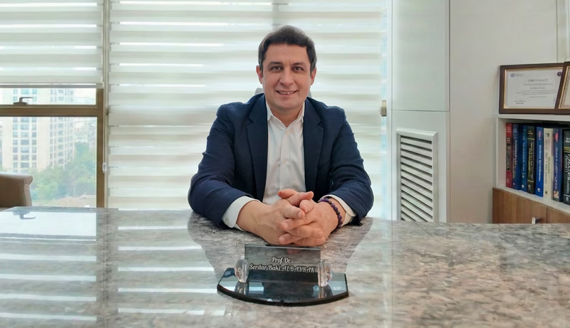
Prof. Dr. Serdar Baki Albayrak
Neurosurgeon · Full Professor of Neurosurgery
Prof. Albayrak performs brain and spinal cord microsurgery with nerve monitoring to protect cord function. For spinal AVMs, he plans each case jointly with an interventional neuroradiology team.
After medical school at Marmara University, he trained in neurosurgery at Istanbul University. Clinical fellowships at the University of Helsinki and Harvard Medical School followed.
27+Years
5,000+Operations
64Countries
33Publications
Figures from Prof. Albayrak's professional profile, September 2026. See his full publication list.
Request an AppointmentFrequently Asked Questions
Can a spinal AVM go away on its own?
No. A spinal AVM or fistula does not close by itself. In fact, a dural fistula usually gets worse over time, so treatment planning should start soon after diagnosis.
Is a spinal AVM life-threatening?
It rarely causes death directly. The main danger is lasting paralysis and bladder loss from a bleed or from congestion. However, a large bleed at neck level can affect breathing.
Is a spinal AVM present from birth?
AVMs inside the cord mostly come from a vessel development problem before birth. In contrast, the common dural fistula develops later in life. Neither type usually runs in families.
How much does spinal AVM surgery cost in Turkey?
The cost depends on the type, the tests you need, surgery or embolization, and the hospital stay. So ask for the cost of your own plan at the consultation, and see our guide to costs.
Prof. Albayrak's Published Research
Prof. Albayrak has no paper on spinal AVMs. The closest work covers vascular microneurosurgery and surgery inside the spinal dura and the spinal cord, the same skills this operation relies on.
Each link opens PubMed; his profile lists all 33 publications.
- Microneurosurgical management of middle cerebral artery bifurcation aneurysms
- A case of intra-dural malignant peripheral nerve sheath tumor in thoracic spine associated with neurofibromatosis type 1
- Cervical leptomeningeal and intramedullary metastasis of a cerebral PNET in an adult
Public health information
- National Institute of Neurological Disorders and Stroke. Arteriovenous Malformations (AVMs).
- MedlinePlus, US National Library of Medicine. Arteriovenous Malformations.
- MedlinePlus. Cerebral angiography (how catheter angiography works).
- NHS. DVT (deep vein thrombosis).
- UK Civil Aviation Authority. Surgical conditions: guidance for health professionals.
- US Centers for Disease Control and Prevention. Medical tourism.
This guide gives general medical information and does not replace a consultation. So diagnosis and treatment decisions need an examination by your own doctor.
Last updated: 29 October 2026. Website editor: Prof. Dr. Serdar Baki Albayrak's clinic, [email protected].
Plan Your Spinal AVM Treatment in Istanbul
Request an appointment with Prof. Dr. Serdar Baki Albayrak. The team then helps you plan the consultation, any angiography and your stay.
Request an Appointment