
Paid online consultations are available by appointment. For booking, please contact us via Whatsapp.

What Is a Brainstem Tumor?
A brainstem tumor is an abnormal growth in the brainstem, the short stalk that links the brain to the spinal cord. This small area controls breathing, swallowing, and eye and face movement. So even a small tumor there can cause clear symptoms.
Most brainstem tumors are gliomas, but they behave very differently. Some grow slowly for years, while others spread quickly through the pons. So the type, more than the place, decides the treatment.
At a Glance
- Where it growsThe midbrain, pons or medulla, at the base of the brain
- Most common typeGliomas, from slow-growing to fast-spreading
- Typical signsDouble vision, a drooping face, poor balance, trouble swallowing
- DiagnosisMRI with contrast; sometimes a biopsy for the exact type
- SurgeryAn option for well-defined tumors near the surface
- Other optionsRadiotherapy, medicines, clinical trials or regular MRI checks
What Brought You Here?
First, pick the line closest to your situation. It then takes you to the matching part of this guide.
- 01Double vision or a face that droopsSigns from the nerves for the eyes and face
- 02Unsteady walking, slurred speech or chokingBalance, speech and swallowing problems
- 03A child with a new squint or frequent fallsHow brainstem tumors show in children
- 04An MRI report mentions a brainstem lesionTumor types and what they mean
- 05You heard the tumor is "inoperable"When surgery helps, and when other treatment comes first
- 06Planning treatment from abroadLength of stay, flights and care back home
Brainstem Tumor Symptoms
Signs often start mildly, so people first blame tiredness or their eyes. However, the brainstem has little spare room, so even a small tumor soon affects several functions.
Double vision and changes in the face
Most nerves for the eyes and face start in the brainstem. So one eye may drift, and double vision follows. Half of the face may also feel numb or droop.
Balance, speech and swallowing
Walking may turn unsteady, and speech slow or slurred. One arm and leg can also weaken. Later, coughing during meals and a hoarse voice point especially to the medulla.
Morning headaches and vomiting
A tumor can also block the flow of spinal fluid and raise the pressure in the skull. Headaches then often feel worst on waking and come with vomiting, MedlinePlus notes. See hydrocephalus treatment for how surgeons relieve it.
Signs in children
Parents may notice a new squint, frequent falls or clumsy hands. In DIPG, these signs appear rapidly, the NCI warns. So a squint with poor balance needs a prompt MRI; see also pediatric neurosurgery.
Where the Tumor Sits Shapes the Symptoms
| Part of the brainstem | What it controls | Typical tumor signs |
|---|---|---|
| Midbrain (top) | Eye movement, pupils, part of the hearing pathway | Double vision, trouble looking up, a drooping eyelid, morning headaches |
| Pons (middle) | Face movement and feeling, chewing, balance links | A weak or numb face, an inward-turning eye, unsteady walking |
| Medulla (bottom) | Breathing, heart rhythm, swallowing, the gag reflex | Choking, a hoarse voice, slurred speech, irregular breathing |
Questions at this stage
What are the first signs of a brainstem tumor?
Often double vision, a drifting eye, a numb or drooping face, or unsteady walking come first. Slurred speech and trouble swallowing may follow. In children, a new squint and frequent falls are common first signs.
When to See a Doctor
Most of these signs build over weeks, so book a prompt visit rather than waiting. Some changes, however, need emergency care the same day.
Book a doctor's visit soon if you notice:
- Double vision or an eye that drifts
- Unsteady walking that keeps getting worse
- Slurred speech, or coughing while you eat
- A child with a new squint, falls or morning vomiting
Seek emergency care for:
- Sudden confusion or deep sleepiness
- Shallow or irregular breathing
- Inability to swallow, or constant choking
- A severe headache with forceful vomiting
- Sudden weakness, numbness or double vision
These signs can mean rising pressure in the skull, a bleed or a stroke. So go to the nearest emergency department first, and plan any trip abroad later.
Types of Brainstem Tumors
Gliomas start in the brain's support cells and make up most brainstem tumors. Yet their behavior differs widely, so the type guides every treatment decision.
| Type | Who it affects | How it grows | Usual approach |
|---|---|---|---|
| Focal glioma | Children and adults | Slowly, with clear edges | Often removable with microsurgery |
| Exophytic glioma | Mostly children | Bulges out of the brainstem | Surgery for the part outside |
| Tectal glioma | Children and young adults | Very slowly, often stable for years | MRI checks; ETV or a shunt for fluid build-up |
| Diffuse midline glioma (DIPG) | Mostly children aged 5 to 10; also adults | Fast, spreading through the pons | Radiotherapy; biopsy in selected cases |
| Adult brainstem glioma | Adults, often aged 30 to 50 | Slow or fast, depending on the grade | Surgery, radiotherapy or monitoring |
| Metastasis | Adults with cancer elsewhere | Spread from another organ | Often radiosurgery |
Focal, Exophytic and Tectal Gliomas
These gliomas usually grow slowly within clear edges, so surgeons can often remove a focal tumor. With an exophytic tumor, the part that bulges outward offers a safer target. A tectal glioma, however, often stays the same size for years, so MRI checks suffice.
Diffuse Midline Glioma and DIPG
These tumors spread through the brainstem without clear edges, and all are grade 4, the NCI notes. DIPG, the form in the pons, mostly affects children aged 5 to 10. Such tumors cannot come out safely, so radiotherapy leads; see also astrocytoma grades.
Brainstem Tumors in Adults
Brainstem gliomas are rare in adults, according to the NCI. Some grow slowly over years, while others are high grade. So the grade decides how much time there is to plan.
NCI experts add that imaging alone cannot confirm a diffuse midline glioma in adults. A biopsy therefore often guides the plan. For tumors elsewhere in the brain, see our overview of brain tumor treatment.
Metastases and Cavernomas
Cancer from the lung, breast, skin or kidney can also spread to the brainstem; see metastatic brain tumors. A cavernoma, by contrast, is a cluster of abnormal blood vessels, not a tumor. Yet its bleeds can mimic a tumor, as our guide to brainstem cavernomas explains.
Are all brainstem tumors cancerous?
No. Tectal and focal low-grade gliomas often grow slowly and may cause few problems for years. However, diffuse midline gliomas and other high-grade tumors are malignant. A cavernoma, too, can look similar but is not a tumor.
How Doctors Diagnose a Brainstem Tumor
First, the neurosurgeon checks your eye movements, face, gag reflex, balance and strength. This exam already points to the affected part of the brainstem. Imaging then confirms the cause.
Prof. Albayrak chooses among these tests, as needed:
- MRI with contrast: the place, size, edges and likely type
- MR spectroscopy: the tumor's chemistry, when a biopsy carries high risk
- Diffusion and perfusion MRI: clues to the grade
- Tractography: nerve pathways, for planning surgery
- Biopsy: the exact type and its gene changes
When a Biopsy Helps
In children, a typical DIPG often shows clearly on MRI alone. In adults, however, several tumors look alike, so a biopsy usually settles the type. It also shows gene changes such as H3 K27M, which some trials require.
Is a biopsy always needed for a brainstem tumor?
Not always, because a typical DIPG in children often shows clearly on MRI. In adults, however, a biopsy usually confirms the type and guides newer medicines. So your surgeon weighs its small risk against what it adds.
Conditions That Can Look Like a Brainstem Tumor
| Condition | Looks similar because | What sets it apart |
|---|---|---|
| Brainstem stroke | Double vision, facial weakness, weak limbs | Starts within minutes; a tumor progresses over weeks |
| Multiple sclerosis | Double vision and poor balance in young adults | Attacks that ease; scattered patches on MRI |
| Brainstem infection or inflammation | Nerve signs that develop quickly | Fever, a recent infection, inflamed spinal fluid |
| Cavernoma bleed | Sudden brainstem symptoms | Traces of old blood on MRI |
| Bell's palsy | Weakness of one side of the face | Only the face nerve; no double vision or poor balance |
| Chiari malformation | Poor balance, swallowing trouble, headaches | A low cerebellum on MRI; no mass |
See also our guide to Chiari malformation. For a mass beside the brainstem, near the hearing nerve, see skull base tumors.
Can a Brainstem Tumor Be Removed?
Sometimes, yes. Surgery suits tumors with clear edges that reach close to the surface of the brainstem. Diffuse tumors, however, mix with healthy nerve tissue, so radiotherapy leads instead.
A deep tumor with no safe route, or a small, slow one, also usually starts with other treatment. So "inoperable" describes one tumor type, not every brainstem tumor.
Can a brainstem tumor be treated without surgery?
Yes, many can. Radiotherapy forms the core of treatment for diffuse tumors such as DIPG. Small, slow tumors may need only MRI checks, and medicines can add to radiotherapy.
Brainstem Tumor Treatment Options Compared
| Option | Who it suits | What it involves | Points to weigh |
|---|---|---|---|
| Microsurgery | Focal and exophytic tumors, some adult gliomas | Removal under the microscope with nerve monitoring | Not possible in diffuse tumors |
| Needle biopsy | An unclear type, or plans for targeted medicines | A small sample through a thin needle, with navigation | Small risk of bleeding or new symptoms |
| Radiotherapy | DIPG and tumors that cannot come out | Daily sessions on weekdays, usually for about six weeks | Often eases symptoms; the effect may fade |
| Radiosurgery | Small, well-defined tumors and metastases | One or a few sessions of focused radiation | Not suitable for large tumors |
| Medicines and clinical trials | High-grade gliomas; known gene changes | Chemotherapy, targeted medicines or trial treatment | Few medicines reach the brainstem well |
| ETV or shunt | Any tumor that blocks spinal fluid | A new route for the fluid, or a drainage tube | Relieves pressure but leaves the tumor |
Brainstem Tumor Surgery: How It Works
Surgery aims to remove as much tumor as is safe while protecting the brainstem. So Prof. Albayrak operates under the microscope, with nerve monitoring throughout.
Safe Entry Points
A few small areas of the brainstem, the safe entry zones, carry fewer vital pathways. First, tractography maps the pathways, and the surgeon then picks the entry closest to the tumor.
Most routes pass through the back of the skull, as in cerebellar tumor surgery.
Nerve Monitoring and Partial Removal
During the operation, monitoring tracks the face nerve, swallowing and the movement pathways. So the surgeon gets a warning as soon as an instrument nears a nerve. If the tumor clings to vital tissue, leaving a thin layer behind protects function.
Biopsy and Relief of Fluid Pressure
A needle biopsy, in contrast, needs only a small opening in the skull. If spinal fluid builds up, an endoscopic third ventriculostomy (ETV) or a shunt also relieves the pressure.
What is the success rate of brainstem tumor surgery?
No single figure applies, because results depend on the tumor type. A focal low-grade glioma that comes out completely often stays controlled for many years. With diffuse tumors, however, surgery cannot remove the tumor, so other treatment leads.
Radiotherapy, Medicines and Oncology Care
Most brainstem tumors need more than one specialist. So Prof. Albayrak plans treatment with radiation and medical oncologists, and also with child cancer specialists for children.
How Radiotherapy Works
Radiotherapy treats tumors that surgery cannot remove. Usually, you receive a few minutes of radiation each weekday for about six weeks. In DIPG, it often eases symptoms for a time.
Doctors also often add steroids to reduce swelling, the NHS notes. Months later, scans may show changes from the radiation itself, so the NCI advises confirming real growth first.
Medicines and Clinical Trials
Chemotherapy and targeted medicines especially help high-grade tumors and those with a known gene change. Still, few medicines reach the brainstem well, so ask early about clinical trials.
Risks of Brainstem Tumor Treatment
Surgery on the brainstem carries real risks, because vital pathways lie close together. Many problems ease over weeks or months, but some can stay.
Possible problems after surgery include:
- New or worse double vision, or facial weakness
- Trouble swallowing, sometimes with a feeding tube for a while
- Poor balance or weakness on one side
- Breathing problems that need support in intensive care
- A spinal fluid leak, infection or fluid build-up
A needle biopsy also carries a small risk of bleeding or new symptoms. Radiotherapy, too, can cause tiredness and, months later, changes in nearby brain tissue.
Is brainstem surgery dangerous?
It carries higher risks than most brain operations, especially for swallowing, the face and the eyes. Nerve monitoring and safe entry points lower these risks. Many new problems also improve with rehabilitation over the following months.
Outlook and Life Expectancy
The outlook depends mostly on the tumor type, and the range is wide. For example, a child with a tectal glioma may live a normal life for years. A diffuse midline glioma, in contrast, remains hard to treat.
| Tumor type | General course | What matters most |
|---|---|---|
| Tectal glioma | Often stable for years, with a normal daily life | Control of fluid build-up |
| Focal low-grade glioma | Long-term control is common after complete removal | How much of the tumor comes out |
| Adult low-grade glioma | Slow change over years | Gene profile and response to treatment |
| High-grade glioma | Months to a few years | Response to radiotherapy and medicines |
| DIPG in children | Most children live less than 2 years after diagnosis (NCI) | Clinical trials and good symptom care |
These figures describe groups, not one person. Research also gives real reasons for hope, especially as gene-based trials grow in DIPG.
Recovery and Follow-Up
After surgery, therapists help with walking, speech and swallowing. An eye doctor can also ease double vision, for example with prism glasses.
Even low-grade tumors can regrow, so Prof. Albayrak usually orders an MRI every three months for two years, then every six months.
What is the life expectancy with a brainstem tumor?
With tectal or focal low-grade gliomas, many people live for many years. In DIPG, however, most children do not live longer than 2 years, the NCI notes. So only your scans and tissue type allow a personal estimate.
Coming to Istanbul from the US, Canada, the UK or Europe
Brainstem treatment needs careful timing, so plan your trip around your recovery. Also keep the return date flexible.
How Long to Stay in Istanbul
First, add up the consultation, the tests, the operation and the hospital stay. Then rest nearby until your check-up. Swallowing or balance problems can also lengthen the stay.
When You Can Fly Home
After surgery inside the skull, UK aviation guidance advises about 7 days before flying. Air left inside the skull can expand at altitude.
The US CDC also warns that flying after surgery raises the risk of blood clots. So your surgeon sets the date after a check-up.
Radiotherapy and Care at Home
Radiotherapy lasts weeks, so many patients have it at home after surgery in Istanbul. For this reason, contact your local oncology team before you travel. Then take home your discharge summary, scan copies and the tissue report in English.
UK drivers should also check the DVLA rules, because brain tumors have their own driving standards. Elsewhere, rules differ, so ask your doctor.
For insurance and Medicare, see brain surgery in Turkey: planning from the US. For NHS rules and travel insurance, see planning from the UK. From Canada, check with your provincial health plan and private insurer before you book.
Your treatment in Istanbul
From Your First Message to Flying Home
Your treatment in Istanbul follows four clear steps. Each step then answers one practical question.
STEP 01
Request an appointment
Use the form on this page, WhatsApp or call +90 532 308 97 72. A short message is enough: your diagnosis, your country and your travel dates.
You can write in English or Turkish, so use whichever feels easier. The team then agrees the consultation date with you.
STEP 02
Consultation and tests in Istanbul
First, Prof. Albayrak examines you at his clinic in Teşvikiye. Next, he goes through your MRI scans with you. If needed, newer scans or tractography can follow in Istanbul.
He then explains which option suits your tumor. Questions worth asking include:
- Does the tumor have clear edges, and is surgery possible?
- Would a biopsy or gene testing change my treatment?
- Is a clinical trial suitable for me?
What should I bring to my brainstem tumor consultation?
First, bring your MRI scans on a disc or USB drive, with the written reports. Also bring any tissue report and notes from earlier treatment. A list of your medicines helps, too.
STEP 03
Surgery or biopsy and hospital stay
If you choose surgery or a biopsy, it takes place in a hospital in Istanbul. Prof. Albayrak names the hospital at the consultation, along with the likely stay.
After major brainstem surgery, the first days usually include intensive care.
STEP 04
Recovery, fitness to fly and follow-up
After discharge, you rest at a nearby hotel, ideally with a companion. Then, before the flight, your surgeon checks you and confirms that you can fly.
Back home, get urgent help for any of these signs:
- A severe or worsening headache, or repeated vomiting
- New trouble swallowing, breathing or speaking
- Fever, or redness, swelling or clear fluid at the wound
- New weakness, double vision or confusion
Can I have radiotherapy and follow-up MRI at home?
Yes, usually with your local oncology team. Take your Istanbul scans, tissue report and discharge summary along. Your team can then plan radiotherapy and compare future scans.
Your Brainstem Tumor Surgeon in Istanbul
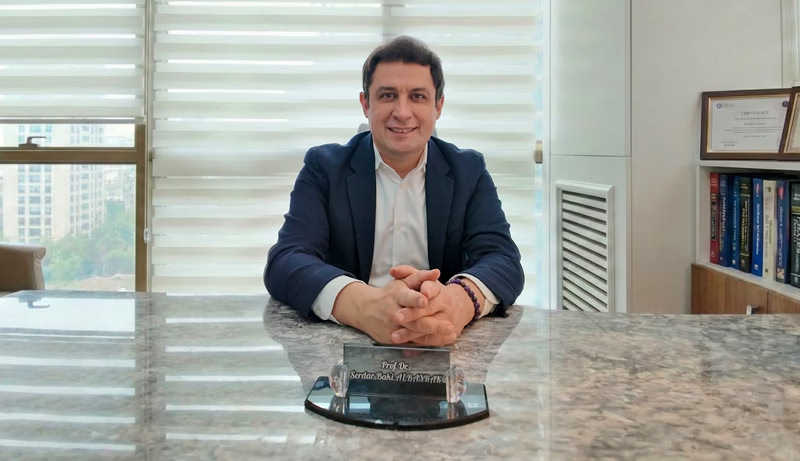
Prof. Dr. Serdar Baki Albayrak
Neurosurgeon · Full Professor of Neurosurgery
In the brainstem, a few millimeters separate a safe route from a lasting deficit. For this reason, Prof. Albayrak plans each approach on MRI and tractography, and he monitors the nerves throughout.
He completed his neurosurgery training at Istanbul University in 2004. He then took clinical fellowships at the University of Helsinki and Harvard Medical School. At Harvard, he trained at Brigham and Women's Hospital.
27+Years
5,000+Operations
64Countries
33Publications
Figures from Prof. Albayrak's professional profile, September 2026. See his full publication list.
Request an AppointmentFrequently Asked Questions
Is a brainstem tumor curable?
Some are: a focal low-grade glioma that comes out completely may never return. Many other tumors, however, need long-term control rather than a cure. So ask what the realistic goal is for your tumor type.
What causes a brainstem tumor?
In most people, no cause appears, and most cases do not run in families. However, neurofibromatosis type 1 and earlier radiation to the head raise the risk. Head injuries and mobile phones have not proven to be causes, MedlinePlus notes.
How much does brainstem tumor surgery cost in Turkey?
The cost depends on the operation, intensive care, the hospital stay and the tests you need. Rehabilitation and your hotel stay also count. So ask for the cost of your own plan at the consultation, before you decide.
Prof. Albayrak's Published Research
One of Prof. Albayrak's first-author reports describes a single metastasis in the medulla that stopped a patient's breathing. Others cover image-guided tumor surgery and the pineal region, which borders the midbrain. Each link opens PubMed; his profile lists all 33 publications.
- Sudden cessation of respiration in a patient with a solitary metastatic focus of renal cell carcinoma in medulla oblongata
- Intra-operative magnetic resonance imaging in neurosurgery
- Microsurgical management of pineal region lesions: personal experience with 119 patients
Public health information
- National Cancer Institute. Childhood Diffuse Intrinsic Pontine Glioma (DIPG).
- National Cancer Institute, NCI-CONNECT. Diffuse Midline Glioma: Diagnosis and Treatment.
- National Cancer Institute. Adult Central Nervous System Tumors Treatment.
- MedlinePlus Medical Encyclopedia, US National Library of Medicine. Brain tumor - primary - adults.
- National Institute of Neurological Disorders and Stroke. Cerebral Cavernous Malformations.
- NHS. Brain tumours.
- UK Civil Aviation Authority. Surgical conditions: guidance for health professionals.
- US Centers for Disease Control and Prevention. Medical tourism.
- GOV.UK, Driver and Vehicle Licensing Agency. Neurological disorders: assessing fitness to drive.
This guide gives general medical information and does not replace a consultation. So diagnosis and treatment decisions need an examination by your own doctor.
Last updated: 1 October 2026. Website editor: Prof. Dr. Serdar Baki Albayrak's clinic, [email protected].
Plan Your Brainstem Tumor Consultation in Istanbul
Request an appointment with Prof. Dr. Serdar Baki Albayrak. The team then helps you plan the dates of your visit.
Request an Appointment